The surgical emergency every new grad fears.
For the non-vets, a "dropped stump" is when a particular artery isn't well tied off when you let go of it (usually accidentally), so uh, blood starts pouring out of it into the abdomen. Pretty much every vet student's nightmare, and inevitable at some point in your career. I've spent years dreading it, being extra super paranoid in every surgery, and it finally happened in August.
Naturally, it was on a day when I was the only surgeon in. And naturally, they had booked a large, deep-chested dog spey for me. It was like looking down into the grand canyon and trying to work with a grappling hook attached to a helicopter. I managed to get my ligatures on, half-blind and up to my elbows in dog abdomen, and, ironically, I wasn't quite confident in them so I wanted to add a third. Just as I lifted the pedicle up to tie another knot, poof, the pedicle was gone. In my hand, securely in the clamp, were the two original ligatures, nice and secure... And attached to nothing.
Sometimes in these situations, blood starts welling up in the abdomen like someone turned on the tap. Fortunately mine didn't bleed all that much, really. I still had to stand there for ten minutes with a fistfull of swabs pressed into the general area, while one of the other vets drove in to help.
At the discharge, I had to figure out how to explain to the client that surgery wasn't exactly routine, but the problem was resolved. I went with "there was more bleeding than usual." At the recheck a week or so later, she mentioned she'd heard a "sloshing sound" the first night. :S
Considering in the very next surgery I did, a cat spey, the clamp cut clean through the uterus and I had to go fishing for the severed end, I was put off surgery for a little while...
Showing posts with label Surgery. Show all posts
Showing posts with label Surgery. Show all posts
Wednesday, 23 December 2015

Wednesday, 9 December 2015
My First Day On The Job Involved A Proptosed Eye
One of the reasons I took the job that I did was that they were interested in hiring a new grad, and willing to give me training wheels for my transition from vet student to actual vet. For my first month, I was never alone in the clinic, so no evenings or weekends, and I was paired with the most experienced senior vet techs. My appointments were all scheduled for 30 minutes instead of twenty, and I was given extra "block off" time for work-ins. My first few days, I didn't have any scheduled appointments at all. I was meant to observe, familiarise myself with where everything is, and maybe jump in on work-ins or walk-ins.
As life would have it, there was a work-in at 8am that first Monday. A dog that had gotten into a fight with its larger friend, and may have gotten a laceration above one eye. "Redness" above the eye, they said. Perfect first case for the new grad, they said.
So in I go and as soon as I lay eyes on the dog, I know exactly what happened, and that I have no idea what to do about it. It wasn't a laceration. It was a proptosed eye.
Small, buggy-eyed dogs like pugs and shih-tzus already have their eyes halfway out of their heads. Therefore, it's pretty easy to pop them the rest of the way out. A solid squeeze or bang on the head could do it. If you catch them straightaway, you can push them right back in. Naturally, this happened the previous afternoon, so the eye had been bulging out exposed to the world for more than 12 hours.
In typical new grad fashion, I gave an excuse to bring the dog out back ("have a look at it under the bright lamp in treatment"), found my boss, and promptly went, "What do I do, what do I do!"
Long story short, we admitted her to the hospital for mini surgery to put the eye back in and suture the eyelids together to hold it. All went well, and I saw her every week for a month or two. The eye retained some function to it and time will tell what happens to it in the long run.
It turns out this was a prelude to the theme of my career. Nothing normal happens to me. It's only the wacky, you-don't-see-that-every-day cases, time and time again.
As life would have it, there was a work-in at 8am that first Monday. A dog that had gotten into a fight with its larger friend, and may have gotten a laceration above one eye. "Redness" above the eye, they said. Perfect first case for the new grad, they said.
So in I go and as soon as I lay eyes on the dog, I know exactly what happened, and that I have no idea what to do about it. It wasn't a laceration. It was a proptosed eye.
Small, buggy-eyed dogs like pugs and shih-tzus already have their eyes halfway out of their heads. Therefore, it's pretty easy to pop them the rest of the way out. A solid squeeze or bang on the head could do it. If you catch them straightaway, you can push them right back in. Naturally, this happened the previous afternoon, so the eye had been bulging out exposed to the world for more than 12 hours.
In typical new grad fashion, I gave an excuse to bring the dog out back ("have a look at it under the bright lamp in treatment"), found my boss, and promptly went, "What do I do, what do I do!"
Long story short, we admitted her to the hospital for mini surgery to put the eye back in and suture the eyelids together to hold it. All went well, and I saw her every week for a month or two. The eye retained some function to it and time will tell what happens to it in the long run.
It turns out this was a prelude to the theme of my career. Nothing normal happens to me. It's only the wacky, you-don't-see-that-every-day cases, time and time again.
Tuesday, 19 August 2014
Horse Anaesthetic Equipment Is Giant-Sized
The title says it all. I scrubbed in on the surgery for my equine case, and as much as I don't like equine surgery, I have to admit, the stuff is pretty cool.
The surgery room itself is big and white and clean feeling, with windows for people outside to look in. It's attached to the recovery box, which is a big black room, and once the horse is induced, they lift it up using a stretcher thingy with giant chains attached to the really high ceiling. The whole wall of the recovery box opens up into surgery, and there's an invisible boundary between the two since surgery is sterile, so a sterile team waits on the other side to take over from the non-sterile team in the recovery box. The horse gets hoisted up, floated around a corner, and set down on the giant surgery table.
The anaesthetic circuit is basically the same as any other circuit, but it's giant-sized. The tubes are as big around as your arm. The ventilator is this giant cylinder. The rebreathing bag is as big as a pillow.
As you might imagine, the fact that horses are so big works against them. If they're on their back, all their organs press against their diaphragm and make it hard to breathe. Their own weight squashes their nerves and muscles and can ruin them if you don't take proper precautions. And that's just the tip of the iceberg of the headache that is equine anaesthesia and surgery.
The surgery room itself is big and white and clean feeling, with windows for people outside to look in. It's attached to the recovery box, which is a big black room, and once the horse is induced, they lift it up using a stretcher thingy with giant chains attached to the really high ceiling. The whole wall of the recovery box opens up into surgery, and there's an invisible boundary between the two since surgery is sterile, so a sterile team waits on the other side to take over from the non-sterile team in the recovery box. The horse gets hoisted up, floated around a corner, and set down on the giant surgery table.
The anaesthetic circuit is basically the same as any other circuit, but it's giant-sized. The tubes are as big around as your arm. The ventilator is this giant cylinder. The rebreathing bag is as big as a pillow.
As you might imagine, the fact that horses are so big works against them. If they're on their back, all their organs press against their diaphragm and make it hard to breathe. Their own weight squashes their nerves and muscles and can ruin them if you don't take proper precautions. And that's just the tip of the iceberg of the headache that is equine anaesthesia and surgery.
Friday, 8 August 2014
Turtle Lamp
You might be wondering how surgeons get inside the turtle. It turns out that one of the methods is to make an incision in the soft area in front of their back legs (or front, depending on what you want to do). Then they can stick an endoscope in, with a light source and a camera on it, and use tiny instruments with long handles. The best part is that the light makes the internal cavity of the turtle glow, and you can see it through the shell on their belly!
I can't find any good photos on google, so I'm going to show you the ones I took. I'm not sure if we're allowed to do that, so uh, don't tell the school on me please.

Intubated and with an IV catheter in her jugular.

The reason the turtle looks so yellow is because there's a sticky sterile covering with iodine in it, called Ioban.

If you look carefully, you can see how bright the pink inside of the turtle is because of the light from the endoscope.
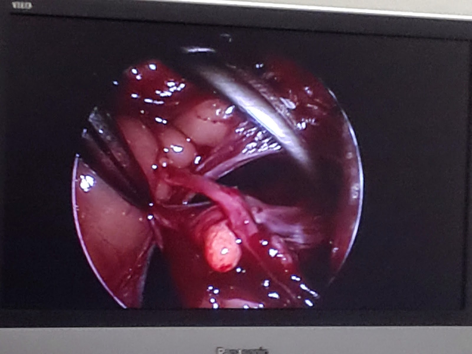
The screen showing the instruments and tissues on the endoscope.
Why was this turtle having surgery? The short and simple version is that it had egg impaction. And how did they definitively diagnose that? A CT scan. In case you were wondering, here's a picture from google of a turtle CT scan:

Thursday, 7 August 2014
We Killed A Dinosaur
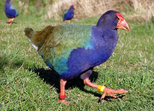
Wildlife medicine in New Zealand is somewhat unique. There are a lot of critters here that don't exist anywhere else, and, conversely, things that aren't here that are everywhere else. During my time on wildbase, I sadly never got to see a snake or ferret. But I did get to see lots of super endangered native creatures, such as takahe. There's like 260 of them left.

And tuatara, which are basically dinosaurs, and the star of today's blog post.

They're basically prehistoric. They're so primitive, they're their own order. I know they look like lizards, but they're closer to dinosaurs. According to wikipedia, "The two species of tuatara are the only surviving members of their order, which flourished around 200 million years ago."
One of them came to the hospital with a diseased eye. It had probably been traumatised (eg poked it into a stick or something) and was now blind, or at least that's my understanding. All their heroic efforts to treat it medically had failed, so it came down to surgery. The first eye enucleation surgery to ever be performed in a tuatara.
The interesting thing about wildlife and exotic medicine is that there isn't a whole mountain of literature for every single species. You might be the second person ever to anaesthetise a giraffe with that combination of drugs, so all you have to go on is one twenty-year old case report and extrapolation from other species. Unfortunately that's not as easy as it sounds, because drugs can have dramatically different effects as species have small differences in enzymes or receptors or whatever. For instance, cats are massively different to dogs in some areas, and we have lots of research about those species. You can imagine how often we diagnose and treat the same disease in, say, a tiger. Now imagine how likely you are to find anything published about a species that numbers in the thousands.
The other fun thing about tuatara surgery is that reptiles are ridiculous animals. The way it was explained to me is that everything in birds happens super fast, they can turn on a dime, and everything that happens in reptiles happens super slow. If a reptile emergency comes in, the first thing you should do is put on the kettle, so you can have a think about it over your tea. Any changes in their health tend to take a long time to manifest. That is important information for this story.
We went ahead with the surgery, which was all quite exciting. It was especially exciting for anaesthesia, who were very stressed out. I don't remember the details because I wasn't on anaesthesia then, but things like heart rate and respiratory rate were apparently quite distressing. So while anaesthesia was freaking out, the wildlife clinician was calmly shrugging it off, with the explanation that some crocodillians can have heart rates as low as one beat per minute. Turtle hearts can keep beating for 24 hours after death (as in, when you open them up at necropsy, you can find their heart beating in front of you... even if you take it out, apparently). Basically, it's impossible to tell if they're dead or alive while under anaesthesia, so he decided not to worry about it. (It fits the theme of not being able to tell if animals are alive while on wildlife roster).
The surgery went along just fine. They packed the empty eye socket with some mini absorbable sponge thingies and sutured it all up. They made it a little bandage that made it look like a pirate. I have an adorable picture, but I doubt I'm supposed to share it on the internet (it might get picked up by google images or something and be stuck there forever when people search about tuatara, and then I think the university might get a teeny bit mad at me).
Details aside, things were fine and dandy that night and into the next morning. During the day, however, the tuatara slowed down, and in grand reptile fashion, we began to question whether it was alive or not. The clinicians broke out more heroics and it went under intensive care for most of the afternoon. Interestingly, since tuatara are in their own order, you need another tuatara if you want to do a blood transfusion... and there's not too many of them around, as you can imagine.
We had the little guy on oxygen, breathing for him, and all sorts of monitoring. He got drugs and fluids and blood products and heating pads and everything we could think of. Unfortunately the monitoring equipment isn't exactly built for tuatara, so the accuracy was questionable, making it even harder to tell anything about the heart or whatever. The heart rate went down and he wouldn't breathe for himself. We did what we could, and gave it a few hours for good measure. He had gone very pale. Surprisingly, we still couldn't figure out if he was actually dead, so we stopped breathing for him and let the chips fall as they may. We put him back in the incubator for the night, just to be sure. The next morinng he hadn't moved any, so we were getting pretty confident we'd lost him.
Too bad for the world's first enucleation on a tuatara. The upside of taking two days to die is that we'd all kind of... accepted it. By the time we gave up on him, I had plenty of time to prepare myself emotionally so I wasn't particularly upset. If he'd given up the ghost in the middle of surgery or something, that would have been more difficult to deal with.
And tuatara, which are basically dinosaurs, and the star of today's blog post.

They're basically prehistoric. They're so primitive, they're their own order. I know they look like lizards, but they're closer to dinosaurs. According to wikipedia, "The two species of tuatara are the only surviving members of their order, which flourished around 200 million years ago."
One of them came to the hospital with a diseased eye. It had probably been traumatised (eg poked it into a stick or something) and was now blind, or at least that's my understanding. All their heroic efforts to treat it medically had failed, so it came down to surgery. The first eye enucleation surgery to ever be performed in a tuatara.
The interesting thing about wildlife and exotic medicine is that there isn't a whole mountain of literature for every single species. You might be the second person ever to anaesthetise a giraffe with that combination of drugs, so all you have to go on is one twenty-year old case report and extrapolation from other species. Unfortunately that's not as easy as it sounds, because drugs can have dramatically different effects as species have small differences in enzymes or receptors or whatever. For instance, cats are massively different to dogs in some areas, and we have lots of research about those species. You can imagine how often we diagnose and treat the same disease in, say, a tiger. Now imagine how likely you are to find anything published about a species that numbers in the thousands.
The other fun thing about tuatara surgery is that reptiles are ridiculous animals. The way it was explained to me is that everything in birds happens super fast, they can turn on a dime, and everything that happens in reptiles happens super slow. If a reptile emergency comes in, the first thing you should do is put on the kettle, so you can have a think about it over your tea. Any changes in their health tend to take a long time to manifest. That is important information for this story.
We went ahead with the surgery, which was all quite exciting. It was especially exciting for anaesthesia, who were very stressed out. I don't remember the details because I wasn't on anaesthesia then, but things like heart rate and respiratory rate were apparently quite distressing. So while anaesthesia was freaking out, the wildlife clinician was calmly shrugging it off, with the explanation that some crocodillians can have heart rates as low as one beat per minute. Turtle hearts can keep beating for 24 hours after death (as in, when you open them up at necropsy, you can find their heart beating in front of you... even if you take it out, apparently). Basically, it's impossible to tell if they're dead or alive while under anaesthesia, so he decided not to worry about it. (It fits the theme of not being able to tell if animals are alive while on wildlife roster).
The surgery went along just fine. They packed the empty eye socket with some mini absorbable sponge thingies and sutured it all up. They made it a little bandage that made it look like a pirate. I have an adorable picture, but I doubt I'm supposed to share it on the internet (it might get picked up by google images or something and be stuck there forever when people search about tuatara, and then I think the university might get a teeny bit mad at me).
Details aside, things were fine and dandy that night and into the next morning. During the day, however, the tuatara slowed down, and in grand reptile fashion, we began to question whether it was alive or not. The clinicians broke out more heroics and it went under intensive care for most of the afternoon. Interestingly, since tuatara are in their own order, you need another tuatara if you want to do a blood transfusion... and there's not too many of them around, as you can imagine.
We had the little guy on oxygen, breathing for him, and all sorts of monitoring. He got drugs and fluids and blood products and heating pads and everything we could think of. Unfortunately the monitoring equipment isn't exactly built for tuatara, so the accuracy was questionable, making it even harder to tell anything about the heart or whatever. The heart rate went down and he wouldn't breathe for himself. We did what we could, and gave it a few hours for good measure. He had gone very pale. Surprisingly, we still couldn't figure out if he was actually dead, so we stopped breathing for him and let the chips fall as they may. We put him back in the incubator for the night, just to be sure. The next morinng he hadn't moved any, so we were getting pretty confident we'd lost him.
Too bad for the world's first enucleation on a tuatara. The upside of taking two days to die is that we'd all kind of... accepted it. By the time we gave up on him, I had plenty of time to prepare myself emotionally so I wasn't particularly upset. If he'd given up the ghost in the middle of surgery or something, that would have been more difficult to deal with.
Monday, 7 April 2014
Turtles and Geckos and Kiwis, Oh My
Day 1 on wildbase was a busy swarm of awesome. The wildlife ward is this tiny room tucked in between the treatment room and the dog kennels in the small animal hospital, which you walk by multiple times a day without ever really noticing. It turns out that just inside, there's a little mini clinic. There's one main room that includes a central surgery area (the whole place isn't much bigger than a dorm room), and then a doorway into the mini wards. There's a large room for a big animal, and another room packed with various sized cages. Five weeks in the smallies hospital, and I never realised there were a bazillion bird patients hanging out right there. Today, there were close to 20 inpatients.
The day started with a bang and never slowed down. On account of being a monday, after a brief tour, I got thrown into the whirlwind of getting food and meds to all 20-odd patients, with only the most basic experience to go on. After cutting up fruits, veggies, and almonds, my first animal-related task was to hold a kiwi while the vet tech force fed it. That's right, a kiwi.

Mondays are apparently particularly hectic because they have rounds on mondays and thursdays. Treatments had to be finished by nine, and then all the wildlife staff (less than ten people) gathered around the tiny clinic to go over the history, diagnostic findings, and treatments for all the patients. There are a number of birds with wing or leg fractures that got surgery, with pins and external fixators and the whole kit and kaboodle. There are a few skinny birds that have been losing weight, a few odds and sods like strange skin lesions or neuro. One bird is in for diagnostics to confirm diabetes insipidus. At the other location, near the large animal teaching unit, there are purportedly three penguins and a few other big birds, but I didn't get out there today. One of the patients is an endangered New Zealand bird, and there are only around 400 left in the world.
After rounds, we had radiology booked for the morning, as we had three birds to radiograph. A harrier, a kingfisher, and some sort of pigeon. Wildbase is great because they have students every week and plunge you right into it. Almost right away, I was positioning the animals, holding, taking blood, making blood smears (badly), running PCV/TP, palpating lesions, all sorts of stuff.

After lunch, there were two consults. One was a sulfur crested cockatoo who we admitted for a whole slew of diagnostics, including bloodwork and skin scrapes. The other, however, made my day. It was a pet turtle with conjunctivitis, secondary to vitamin A deficiency (quite a common deficiency in exotic pets). So we did an ophthalmic exam on this turtle. I got to hold it while we examined and treated this turtle, and turtles are exactly as cute and awesome as you'd expect them to be (as long as you don't get bitten). Its little legs waggled around as I sandwiched it between my palms.

After the consults, it was time for the gecko surgery. Yeah, gecko surgery. It had a mass on its neck that was removed. Anaesthetists joined us to manage the anaesthesia, and the whole process of induction and intubation was quite an undertaking. The gecko was on a mask, and when it was asleep enough to lose its "righting reflex" when flipped over, they tried to get the endotracheal tube in. However, the gas anaesthetic diffuses out of the lungs pretty quickly, so there was a limited amount of time for each attempt. Because of the unusual anatomy, it took many attempts.
Despite all that, overall, the procedure wasn't much different than it would be for any other animal. The concerns about respiration and blood pressure are basically the same, it's just a tiny-sized animal. Surgical technique is the same. Suturing is the same, it's just scaly reptilian skin instead. Though, there are a lot of quirks to bear in mind as well. For instance, turtles' lungs are attached to the dorsal carapace, so if you need to ventilate them, you can flap their legs back and forth, and it actually works the lungs!
I also learned that the bottoms of geckos' feet are really, really cool. Not only do they look neat, they feel pretty neat, too.

The day started with a bang and never slowed down. On account of being a monday, after a brief tour, I got thrown into the whirlwind of getting food and meds to all 20-odd patients, with only the most basic experience to go on. After cutting up fruits, veggies, and almonds, my first animal-related task was to hold a kiwi while the vet tech force fed it. That's right, a kiwi.
One of the patients is a Kaka.
Mondays are apparently particularly hectic because they have rounds on mondays and thursdays. Treatments had to be finished by nine, and then all the wildlife staff (less than ten people) gathered around the tiny clinic to go over the history, diagnostic findings, and treatments for all the patients. There are a number of birds with wing or leg fractures that got surgery, with pins and external fixators and the whole kit and kaboodle. There are a few skinny birds that have been losing weight, a few odds and sods like strange skin lesions or neuro. One bird is in for diagnostics to confirm diabetes insipidus. At the other location, near the large animal teaching unit, there are purportedly three penguins and a few other big birds, but I didn't get out there today. One of the patients is an endangered New Zealand bird, and there are only around 400 left in the world.
After rounds, we had radiology booked for the morning, as we had three birds to radiograph. A harrier, a kingfisher, and some sort of pigeon. Wildbase is great because they have students every week and plunge you right into it. Almost right away, I was positioning the animals, holding, taking blood, making blood smears (badly), running PCV/TP, palpating lesions, all sorts of stuff.
A harrier.
After lunch, there were two consults. One was a sulfur crested cockatoo who we admitted for a whole slew of diagnostics, including bloodwork and skin scrapes. The other, however, made my day. It was a pet turtle with conjunctivitis, secondary to vitamin A deficiency (quite a common deficiency in exotic pets). So we did an ophthalmic exam on this turtle. I got to hold it while we examined and treated this turtle, and turtles are exactly as cute and awesome as you'd expect them to be (as long as you don't get bitten). Its little legs waggled around as I sandwiched it between my palms.
Ours was larger, but looked basically the same.
After the consults, it was time for the gecko surgery. Yeah, gecko surgery. It had a mass on its neck that was removed. Anaesthetists joined us to manage the anaesthesia, and the whole process of induction and intubation was quite an undertaking. The gecko was on a mask, and when it was asleep enough to lose its "righting reflex" when flipped over, they tried to get the endotracheal tube in. However, the gas anaesthetic diffuses out of the lungs pretty quickly, so there was a limited amount of time for each attempt. Because of the unusual anatomy, it took many attempts.
Despite all that, overall, the procedure wasn't much different than it would be for any other animal. The concerns about respiration and blood pressure are basically the same, it's just a tiny-sized animal. Surgical technique is the same. Suturing is the same, it's just scaly reptilian skin instead. Though, there are a lot of quirks to bear in mind as well. For instance, turtles' lungs are attached to the dorsal carapace, so if you need to ventilate them, you can flap their legs back and forth, and it actually works the lungs!
I also learned that the bottoms of geckos' feet are really, really cool. Not only do they look neat, they feel pretty neat, too.
More surgeries and radiographs and anaesthetics on the schedule for tomorrow, with all sorts of native New Zealand birds. Should be pretty exciting!
Friday, 24 January 2014
Said in Surgery Rounds
Surgeon: What's a "cherry eye"?
Me: The gland of the third eyelid is prolapsed.
Surgeon: No. What's the gland called?
Students: Nictitating gland?
Surgeon: Yes. And what's wrong with it?
...
Surgeon: It's PROLAPSED
Surgeon: What's a type I open fracture mean? What does the fracture do?
Student: Do?
Surgeon: Where does the bone go?
Student: Uh...?
Surgeon: From the inside to the outside!
Student: How else would the bone go?!
Surgeon: What's the third type of plate?
Students: It makes a bridge.
Surgeon: Yes but what's it called?
Students: A... bridge plate?
*Surgeon starts drawing*
Surgeon: Picture Notre Dam. These things off to the side that support it.
*Draws big arches*
Students: Arches? Scaffold?
*Surgeon writes B _ _ _ _ _ _ _*
Students: Bridge! Bulwark! A! E! O!
*Head shaking*
Students: U!
*Writes B U _ _ _ _ S S*
Students: Buttress!
Surgeon: What type of screws would you use?
Student: Small ones.
Me: The gland of the third eyelid is prolapsed.
Surgeon: No. What's the gland called?
Students: Nictitating gland?
Surgeon: Yes. And what's wrong with it?
...
Surgeon: It's PROLAPSED
Surgeon: What's a type I open fracture mean? What does the fracture do?
Student: Do?
Surgeon: Where does the bone go?
Student: Uh...?
Surgeon: From the inside to the outside!
Student: How else would the bone go?!
Surgeon: What's the third type of plate?
Students: It makes a bridge.
Surgeon: Yes but what's it called?
Students: A... bridge plate?
*Surgeon starts drawing*
Surgeon: Picture Notre Dam. These things off to the side that support it.
*Draws big arches*
Students: Arches? Scaffold?
*Surgeon writes B _ _ _ _ _ _ _*
Students: Bridge! Bulwark! A! E! O!
*Head shaking*
Students: U!
*Writes B U _ _ _ _ S S*
Students: Buttress!
Surgeon: What type of screws would you use?
Student: Small ones.
Friday, 17 January 2014
Until Blood Started Mysteriously Pooling Out of Nowhere
The curriculum's vision of "spey class" doesn't align well with the reality.
1. There are often not enough speys in a week for all four students.
2. People seem to cancel these appointments at an alarming rate.
3. The animals we do get aren't exactly young and healthy.
Earlier in the week, my classmate's spey was some several hour ordeal that involved many stacks of blood-soaked gauze and a uterus as thick as hose pipe. My spey got cancelled completely because of a UTI. My friend shared her spey with me, which turned out to be a 4-year-old shelter dog that came off of heat a week ago.
Now, when they're in heat, their uterus gets huge and very vascular. This complicates things because the usually teeny vessels become a lot bigger and you have to worry more about tying them all off, and as you can imagine, a big hose pipe is harder to clamp off than a small one. Despite this, everything was going mostly hunky-dory--at least, until blood started mysteriously pooling out of nowhere.
It started as a general oozing, an unusual amount of fluid down in the body cavity. Gradually, the pinky-orangey fluid deep down turned into really-quite-red fluid all over my side of the dog.
When you do a spey, you tie off the vessels that connect the ovary to the body, then cut the ovary off; this leaves you with a pedicle of tissue and tied-off vessels that you drop back into the body. You check this to make sure you really got the vessels all closed off, or else they just keep bleeding forever. If you accidentally drop the pedicle before you're ready, or the ligature is too loose and comes off, it's called a "dropped stump" and is what gives fledgling vet students night terrors. No matter how many times your professors explain the steps to find and retrieve your dropped stump, it's still a tiny fleshy blob in the middle of a bunch of other fleshy blobs, all submerged beneath an ocean of blood with a rapidly rising tide. It hasn't happened to me, but I saw a vet deal with it once.
This isn't what happened today, but now you can understand why I became alarmed that my severed pedicle was suddenly sitting in a pool of blood. The vet teaching us took some time to check out the stump, search around in the body cavity, and couldn't find the source, so he was like, "Nevermind that, we'll worry about it later," and my friend finished her ovary and we went on to the uterine body.
As time went on, the oozing-become-pooling didn't subside, and the vet kept checking but then deciding we'd "worry about it later." Since he couldn't figure out what was going on (my stump wasn't what was bleeding, phew), he had the nurses page the surgical resident. She joined us, and even she couldn't figure out where all the blood was coming from. They hooked up suction and diathermy, packed the abdomen with laparotomy sponges, and threw in a whole bunch of ligatures onto various fleshy blobs, but it didn't seem to change much. The blood was coming from everywhere and nowhere.
Eventually, they got it from "pooling" back to "oozing" and decided to close up the wound, but when discussing medications with me, made the point that she could require a second emergency surgery if it turned out she kept bleeding on the inside. Fortunately there are a bazillion people in the ICU whose job it is to keep an eye on patients like this one. However, as four hours of sore feet can attest, the vet made the very good point that dogs on heat are not very good cases for student spey classes.
1. There are often not enough speys in a week for all four students.
2. People seem to cancel these appointments at an alarming rate.
3. The animals we do get aren't exactly young and healthy.
Earlier in the week, my classmate's spey was some several hour ordeal that involved many stacks of blood-soaked gauze and a uterus as thick as hose pipe. My spey got cancelled completely because of a UTI. My friend shared her spey with me, which turned out to be a 4-year-old shelter dog that came off of heat a week ago.
Now, when they're in heat, their uterus gets huge and very vascular. This complicates things because the usually teeny vessels become a lot bigger and you have to worry more about tying them all off, and as you can imagine, a big hose pipe is harder to clamp off than a small one. Despite this, everything was going mostly hunky-dory--at least, until blood started mysteriously pooling out of nowhere.
It started as a general oozing, an unusual amount of fluid down in the body cavity. Gradually, the pinky-orangey fluid deep down turned into really-quite-red fluid all over my side of the dog.
When you do a spey, you tie off the vessels that connect the ovary to the body, then cut the ovary off; this leaves you with a pedicle of tissue and tied-off vessels that you drop back into the body. You check this to make sure you really got the vessels all closed off, or else they just keep bleeding forever. If you accidentally drop the pedicle before you're ready, or the ligature is too loose and comes off, it's called a "dropped stump" and is what gives fledgling vet students night terrors. No matter how many times your professors explain the steps to find and retrieve your dropped stump, it's still a tiny fleshy blob in the middle of a bunch of other fleshy blobs, all submerged beneath an ocean of blood with a rapidly rising tide. It hasn't happened to me, but I saw a vet deal with it once.
This isn't what happened today, but now you can understand why I became alarmed that my severed pedicle was suddenly sitting in a pool of blood. The vet teaching us took some time to check out the stump, search around in the body cavity, and couldn't find the source, so he was like, "Nevermind that, we'll worry about it later," and my friend finished her ovary and we went on to the uterine body.
As time went on, the oozing-become-pooling didn't subside, and the vet kept checking but then deciding we'd "worry about it later." Since he couldn't figure out what was going on (my stump wasn't what was bleeding, phew), he had the nurses page the surgical resident. She joined us, and even she couldn't figure out where all the blood was coming from. They hooked up suction and diathermy, packed the abdomen with laparotomy sponges, and threw in a whole bunch of ligatures onto various fleshy blobs, but it didn't seem to change much. The blood was coming from everywhere and nowhere.
Eventually, they got it from "pooling" back to "oozing" and decided to close up the wound, but when discussing medications with me, made the point that she could require a second emergency surgery if it turned out she kept bleeding on the inside. Fortunately there are a bazillion people in the ICU whose job it is to keep an eye on patients like this one. However, as four hours of sore feet can attest, the vet made the very good point that dogs on heat are not very good cases for student spey classes.
Wednesday, 15 January 2014
The Longest Day Ever
Three posts in one day, what is this?! So much happened, so many stories!
I'm going to admit a secret. I'm not actually writing this on wednesday. As you can imagine, three posts' worth of stories also meant I was completely exhausted that day. It may be a blogging faux pas, but I quite often write posts and sneak them into the archives 1984-style. I guess it's because the blog also functions as a diary and I like to keep things in a mostly chronological order. It's very likely that posts will continue to pop up "in the past" as I write them and then pretend nothing's changed.
I almost didn't write this one, because I already told the major "stories" (my wuss of a patient and the rabbit castration) and this is going to be more rambly. However, I decided I couldn't not mention my first real surgery.
Somehow, I went from having zero patients, to one medicine patient (that I kind of commandeered from the medicine student) and two surgeries. One of my roster-mates was scheduled to do both a spey and a castration today, so she suggested trading her castration today for my castration on friday. This was after the clinician offered the rabbit castration to me, so I accepted both quite happily. I also wanted to stick with my original patient and find out what the results of her urinalysis were, which naturally led into me also doing up her meds and discharge.
The castration was interesting because the dog is participating in a study looking at the effects of bupivicaine, a local anaesthetic, in the injection site. He was one of the controls, so didn't get any bupivicaine, but he still got hooked up to an EEG all during the surgery. I also got to work with a vet that I haven't seen before or since, who turned out to be a really nice guy and an amazing help. My friend did her surgery with him as well, and we agreed he had the perfect balance of explaining what to do, but allowing us to actually do it ourselves. He was thorough, patient, and supportive. (To be fair, almost all the vets are like that, except for one surgeon that I have been calling eats-students-for-breakfast in previous posts). But this guy is mysterious because, like I mentioned, no one has seen him around the hospital except for that one day.
The reason I called this the longest day ever isn't because of the actual hour total. Last week on referral surgery I had a few 12-14 hour days. But this day was so exhausting. I did my dog castration, my rabbit castration, chased a vet all over the hospital to talk about all three of my patients--including what meds to do and for how long, specifics on the discharge information and home advice, and writing up the surgery report. What really sent me over the edge was a big practical lab in the afternoon, about dentistry.
These labs apparently happen every wednesday afternoon and are mandatory. As usual, I was late, because I had to do all those things for all my patients. The lab started at around 4pm, and I had been standing and moving around all day, so I was already tired. I don't find dentistry particularly exciting, and this lab was exactly the same as the lab in fourth year (severed heads and all), our dead dogs a few weeks ago, and the actual dentals we did on monday. Clean teeth, extract teeth, queue up for the drill. Zzzzzzz. Ten or fifteen minutes into it, I was sagging on my stool, while the person next to me kept nodding off. Important as it is, a lab like this really needs to be in the morning, not 4pm. I ran off to discharge my rabbit patient before I completely fell asleep.
I'm going to admit a secret. I'm not actually writing this on wednesday. As you can imagine, three posts' worth of stories also meant I was completely exhausted that day. It may be a blogging faux pas, but I quite often write posts and sneak them into the archives 1984-style. I guess it's because the blog also functions as a diary and I like to keep things in a mostly chronological order. It's very likely that posts will continue to pop up "in the past" as I write them and then pretend nothing's changed.
I almost didn't write this one, because I already told the major "stories" (my wuss of a patient and the rabbit castration) and this is going to be more rambly. However, I decided I couldn't not mention my first real surgery.
Somehow, I went from having zero patients, to one medicine patient (that I kind of commandeered from the medicine student) and two surgeries. One of my roster-mates was scheduled to do both a spey and a castration today, so she suggested trading her castration today for my castration on friday. This was after the clinician offered the rabbit castration to me, so I accepted both quite happily. I also wanted to stick with my original patient and find out what the results of her urinalysis were, which naturally led into me also doing up her meds and discharge.
The castration was interesting because the dog is participating in a study looking at the effects of bupivicaine, a local anaesthetic, in the injection site. He was one of the controls, so didn't get any bupivicaine, but he still got hooked up to an EEG all during the surgery. I also got to work with a vet that I haven't seen before or since, who turned out to be a really nice guy and an amazing help. My friend did her surgery with him as well, and we agreed he had the perfect balance of explaining what to do, but allowing us to actually do it ourselves. He was thorough, patient, and supportive. (To be fair, almost all the vets are like that, except for one surgeon that I have been calling eats-students-for-breakfast in previous posts). But this guy is mysterious because, like I mentioned, no one has seen him around the hospital except for that one day.
The reason I called this the longest day ever isn't because of the actual hour total. Last week on referral surgery I had a few 12-14 hour days. But this day was so exhausting. I did my dog castration, my rabbit castration, chased a vet all over the hospital to talk about all three of my patients--including what meds to do and for how long, specifics on the discharge information and home advice, and writing up the surgery report. What really sent me over the edge was a big practical lab in the afternoon, about dentistry.
These labs apparently happen every wednesday afternoon and are mandatory. As usual, I was late, because I had to do all those things for all my patients. The lab started at around 4pm, and I had been standing and moving around all day, so I was already tired. I don't find dentistry particularly exciting, and this lab was exactly the same as the lab in fourth year (severed heads and all), our dead dogs a few weeks ago, and the actual dentals we did on monday. Clean teeth, extract teeth, queue up for the drill. Zzzzzzz. Ten or fifteen minutes into it, I was sagging on my stool, while the person next to me kept nodding off. Important as it is, a lab like this really needs to be in the morning, not 4pm. I ran off to discharge my rabbit patient before I completely fell asleep.
First Solo Surgery Was A Rabbit Castration
Since my spey today was cancelled, the vet running our roster suggested I scrub in for the rabbit coming in. I saw one last week; the student had scrubbed in, but the vet did the surgery. Thus I was not expecting to be the sole surgeon on this rabbit.
After anaesthesia got the rabbit asleep, I went ahead and scrubbed and gowned. I didn't think it was strange that the vet hadn't scrubbed yet, because they often wander around doing other things and scrub in quickly at the last minute. However, as I stand there awkwardly with my hands clasped in front of me, peeking into the anaesthesia room, the vet turns to me and says, "Okay, come in in and we'll get started. You can go ahead and drape." Que question marks over my head--isn't she scrubbing in, too?
No. It was just me. She directed me, but I was the only person scrubbed in for the first time ever, and it was a rabbit. Rabbit castrations are done just in the anaesthesia room, rather than the operating theatre, which is quite a tiny space. I guess that is fitting for quite a tiny patient. We set up my instruments just on a stool, and I was given the warning, "Don't knock this over," as the stool seat is exactly the same width as the box holding the instruments. I also had an awkward time draping, since the table was up against a wall--how do you get around the other side? I ended up doing three drapes in a triangle rather than four in a square.
And then we were off. Rabbit balls are long and kidney-bean shaped, and they also sometimes get sucked back into the abdomen. This happened as I was working on the first one, and the anaesthesia student had to reach under the drapes and press on the abdomen to pop the testicle back out. Another difference is that they are actually adhered to the skin, so you have to dissect them free (and not drop them into the abdomen while doing it). But overall, it was fairly simple, and I got out both balls without a hitch.
My only mistake is that I'm not used to working on live animals, and no one has really taught us about tissue handling, so I kept clamping the instruments on too tight. You see, I have haemostats at home to practice with, and I just clamp and unclamp them, plus they are difficult to operate so I got good at squeezing them really hard. Even in labs it's never mattered whether you do one, two, or three clicks. So I just squeeze my hand and get whatever amount of clicks I end up getting, without really thinking about it. But as I learned, three clicks is way too many for poor, delicate rabbit skin and tissues, affirmed by the vet's repeated wincing.
After anaesthesia got the rabbit asleep, I went ahead and scrubbed and gowned. I didn't think it was strange that the vet hadn't scrubbed yet, because they often wander around doing other things and scrub in quickly at the last minute. However, as I stand there awkwardly with my hands clasped in front of me, peeking into the anaesthesia room, the vet turns to me and says, "Okay, come in in and we'll get started. You can go ahead and drape." Que question marks over my head--isn't she scrubbing in, too?
No. It was just me. She directed me, but I was the only person scrubbed in for the first time ever, and it was a rabbit. Rabbit castrations are done just in the anaesthesia room, rather than the operating theatre, which is quite a tiny space. I guess that is fitting for quite a tiny patient. We set up my instruments just on a stool, and I was given the warning, "Don't knock this over," as the stool seat is exactly the same width as the box holding the instruments. I also had an awkward time draping, since the table was up against a wall--how do you get around the other side? I ended up doing three drapes in a triangle rather than four in a square.
And then we were off. Rabbit balls are long and kidney-bean shaped, and they also sometimes get sucked back into the abdomen. This happened as I was working on the first one, and the anaesthesia student had to reach under the drapes and press on the abdomen to pop the testicle back out. Another difference is that they are actually adhered to the skin, so you have to dissect them free (and not drop them into the abdomen while doing it). But overall, it was fairly simple, and I got out both balls without a hitch.
My only mistake is that I'm not used to working on live animals, and no one has really taught us about tissue handling, so I kept clamping the instruments on too tight. You see, I have haemostats at home to practice with, and I just clamp and unclamp them, plus they are difficult to operate so I got good at squeezing them really hard. Even in labs it's never mattered whether you do one, two, or three clicks. So I just squeeze my hand and get whatever amount of clicks I end up getting, without really thinking about it. But as I learned, three clicks is way too many for poor, delicate rabbit skin and tissues, affirmed by the vet's repeated wincing.
Friday, 10 January 2014
The Harrowing Tale of Hip Fracture Friday
This morning was rather stressful because apparently all the instructions for today were given at afternoon rounds yesterday, which I missed because I was in surgery. All the cases for today had been divvied up, leaving me with no options.
The nurse put my name down for a lame labrador. It seemed fairly interesting, so I didn't mind too much. About five minutes later, another student approaches me and tells me he'd actually claimed that case last night, but didn't put his name into the system. I find out later that this student was supposed to find me after rounds yesterday and talk about the remaining cases, find out which one I wanted and sort out what was going to happen this morning. He didn't do that. He picked a case and went home.
At first, I put up a defense. "I haven't had a chance to do an orthopaedic exam yet," I explained, basically thinking you should have put your damn name on it, it's mine now. He agreed he would just come in on the consult and we would share the case. I go off and read all the patient notes, and eventually learn that he's started studying up on a completely different case and I may have successfully stolen this case for myself. I was a bit relieved, because it's unfair that my day would have been entirely screwed up and it was all his fault for not finding me yesterday afternoon while I was in theatre.
Then, as I'm reading the patient notes, a popup says one of my classmates is looking at that file on another computer. He's not on surgery this week, so I go ask him what's up, and he says it's a medicine case. We scratch our heads over this until we figure out the consult is scheduled under both medicine and surgery. I investigate, and long story short, it's a medicine case. No more case for me.
Just as I was about to get stressed out again, a new, urgent case pops up. A puppy fractured its femur. A case! Turns out it was not really the best case to have on a friday.
I did the consult when they arrived, and was pleased to find that I'm slightly less bumbly than I was in the first one. I was also pleased to find that the clients were willing to pay for the expensive fancy surgery. Thus the patient came under my care and dominated my entire day. Mostly that meant setting up a cage for him and then doing tons of paperwork.
In the afternoon, we went into surgery. It was interesting, but the hip area is challenging and the surgeon had a difficult time pinning the fracture back together and making sure he got all the pieces in the correct position. I became very glad that I was with the chillax surgeon, because eats-students-for-breakfast tends to get frustrated quickly and start throwing instruments around the room (not even joking). This guy was basically like "Aw, damn it," in a completely relaxed manner as he casually mentions he can't see anything and has no idea if his pins are in the correct place. After three hours of digging around in the hip joint trying to reduce the fracture and hold it together, he sews up a bazillion muscles, closes the wound, and we're off to radiology. However, if the pins aren't in the correct place, we have to come back in, take everything out, and do the alternate procedure where we just remove the femoral head rather than trying to hold it together with pins.
Guess what we found on xrays?
The sad thing is that the femoral neck, our goal during surgery, was very nicely aligned. He had fixed that part of the fracture perfectly. It just turns out that there was another fragment that must have come from somewhere else. Therefore, we had to go back in, re-scrub, undo all the skin staples and all the sutured muscles, and dive back down to the joint. At this point it's 6pm and we're looking at probably 2-4 more hours.
Before committing to the femoral head osteotomy, he tried to find that extra fragment, but couldn't. As far as he could tell, the joint surface was undamaged, though it was possible the fragment was too deep for us to reach. He called in the other surgeon, because once you take out the femoral head there's no going back--there's no chance of future improvements if the patient doesn't do well, like you can't do a total hip replacement when the puppy gets older. So we wait around for the other surgeon to arrive, and then they peer at it and poke at it for a good twenty minutes, discussing the pros and cons and what they think about what they see. The entire time, I'm thinking leave it, leave it, leave it. As much as I would like to see an FHO performed, I really didn't want to be there until 10pm on a friday. Finally, after much deliberation, they decided the best standard of care would be to leave the repair in place, and come back for a second operation if an FHO becomes necessary in the future. Phew!
I still had to write a surgery report, sort out some extra post-op meds, and do all the puppy's paperwork for the weekend, but at least I got home while it was still light out.
The nurse put my name down for a lame labrador. It seemed fairly interesting, so I didn't mind too much. About five minutes later, another student approaches me and tells me he'd actually claimed that case last night, but didn't put his name into the system. I find out later that this student was supposed to find me after rounds yesterday and talk about the remaining cases, find out which one I wanted and sort out what was going to happen this morning. He didn't do that. He picked a case and went home.
At first, I put up a defense. "I haven't had a chance to do an orthopaedic exam yet," I explained, basically thinking you should have put your damn name on it, it's mine now. He agreed he would just come in on the consult and we would share the case. I go off and read all the patient notes, and eventually learn that he's started studying up on a completely different case and I may have successfully stolen this case for myself. I was a bit relieved, because it's unfair that my day would have been entirely screwed up and it was all his fault for not finding me yesterday afternoon while I was in theatre.
Then, as I'm reading the patient notes, a popup says one of my classmates is looking at that file on another computer. He's not on surgery this week, so I go ask him what's up, and he says it's a medicine case. We scratch our heads over this until we figure out the consult is scheduled under both medicine and surgery. I investigate, and long story short, it's a medicine case. No more case for me.
Just as I was about to get stressed out again, a new, urgent case pops up. A puppy fractured its femur. A case! Turns out it was not really the best case to have on a friday.
I did the consult when they arrived, and was pleased to find that I'm slightly less bumbly than I was in the first one. I was also pleased to find that the clients were willing to pay for the expensive fancy surgery. Thus the patient came under my care and dominated my entire day. Mostly that meant setting up a cage for him and then doing tons of paperwork.
In the afternoon, we went into surgery. It was interesting, but the hip area is challenging and the surgeon had a difficult time pinning the fracture back together and making sure he got all the pieces in the correct position. I became very glad that I was with the chillax surgeon, because eats-students-for-breakfast tends to get frustrated quickly and start throwing instruments around the room (not even joking). This guy was basically like "Aw, damn it," in a completely relaxed manner as he casually mentions he can't see anything and has no idea if his pins are in the correct place. After three hours of digging around in the hip joint trying to reduce the fracture and hold it together, he sews up a bazillion muscles, closes the wound, and we're off to radiology. However, if the pins aren't in the correct place, we have to come back in, take everything out, and do the alternate procedure where we just remove the femoral head rather than trying to hold it together with pins.
Guess what we found on xrays?
The sad thing is that the femoral neck, our goal during surgery, was very nicely aligned. He had fixed that part of the fracture perfectly. It just turns out that there was another fragment that must have come from somewhere else. Therefore, we had to go back in, re-scrub, undo all the skin staples and all the sutured muscles, and dive back down to the joint. At this point it's 6pm and we're looking at probably 2-4 more hours.
Before committing to the femoral head osteotomy, he tried to find that extra fragment, but couldn't. As far as he could tell, the joint surface was undamaged, though it was possible the fragment was too deep for us to reach. He called in the other surgeon, because once you take out the femoral head there's no going back--there's no chance of future improvements if the patient doesn't do well, like you can't do a total hip replacement when the puppy gets older. So we wait around for the other surgeon to arrive, and then they peer at it and poke at it for a good twenty minutes, discussing the pros and cons and what they think about what they see. The entire time, I'm thinking leave it, leave it, leave it. As much as I would like to see an FHO performed, I really didn't want to be there until 10pm on a friday. Finally, after much deliberation, they decided the best standard of care would be to leave the repair in place, and come back for a second operation if an FHO becomes necessary in the future. Phew!
I still had to write a surgery report, sort out some extra post-op meds, and do all the puppy's paperwork for the weekend, but at least I got home while it was still light out.
Thursday, 9 January 2014
Wrist Day
Since all my other patients have gone home, today was predominated by wrist-cat. This sweet little girl was found limping and it turns out she destroyed the ligaments in one of her wrists, probably from jumping down a big height and landing too hard on it. This is the same cat I admitted yesterday and we took more x-rays yesterday afternoon, confirming the need for surgery today.
The surgery is a salvage procedure where we fuse the carpal joints together so they don't end up becoming all degenerated and painful due to the instability from the ligaments. It was pretty much the coolest surgery I've seen. We opened up the wrist, destroyed all the cartilage with a burr, harvested bone from the shoulder and put a bone graft into the wrist, and then put on this absolutely tiny plate with even tinier screws. The idea is that the joints all heal into a big solid stretch of bone that is no longer flexible (fused into a normal standing position so she can still walk normally). There was lots of drilling and gadgets and metal implants and it was all very exciting. It was also my first time working with the really chillax surgeon. You see, there are two surgeons, one who eats students for breakfast, and one who jokes around and is very pleasant.
Unfortunately I missed afternoon rounds, because the surgery was long and started about they same time they did. This was slightly defeating because on monday, eats-students-for-breakfast gave us homework to present on thursday rounds, and all week I'd struggled to fit in my research and make notes. Turns out it didn't matter at all.
While I was waiting for the afternoon surgery, I adopted a fun patient that came in for an emergency Caesarian last night, and now had three brand new puppies. I watched over her, we did a bit of bloodwork, then I did the discharge with the client. She was a sweet little white fluffy dog, and her puppies were so tiny and silly and adorable.
In other news, we had a communications tutorial yesterday afternoon, talking about why clients leave practises and some common mistakes vets and vet students make. The lady shared some funny stories with us about students calling up the wrong client, and giving them a wonderful update about an animal that isn't theirs. She says there are mistakes every year: every year, animals get overdosed because of decimal places being in the wrong place, subcutaneous injections get given IV, forms get messed up and things don't go through. One time, a form didn't go through for an animal to get cremated, and this vet had seriously debated whether they should give the client some random ashes and pretend. They didn't actually do that, and she was very glad they admitted their mistake, because the first thing the client said was "I'm so glad you didn't try to pass off some other animal's ashes!" The vet said she was glad that conversation happened over the phone, because her face went bright red.
The surgery is a salvage procedure where we fuse the carpal joints together so they don't end up becoming all degenerated and painful due to the instability from the ligaments. It was pretty much the coolest surgery I've seen. We opened up the wrist, destroyed all the cartilage with a burr, harvested bone from the shoulder and put a bone graft into the wrist, and then put on this absolutely tiny plate with even tinier screws. The idea is that the joints all heal into a big solid stretch of bone that is no longer flexible (fused into a normal standing position so she can still walk normally). There was lots of drilling and gadgets and metal implants and it was all very exciting. It was also my first time working with the really chillax surgeon. You see, there are two surgeons, one who eats students for breakfast, and one who jokes around and is very pleasant.
Unfortunately I missed afternoon rounds, because the surgery was long and started about they same time they did. This was slightly defeating because on monday, eats-students-for-breakfast gave us homework to present on thursday rounds, and all week I'd struggled to fit in my research and make notes. Turns out it didn't matter at all.
While I was waiting for the afternoon surgery, I adopted a fun patient that came in for an emergency Caesarian last night, and now had three brand new puppies. I watched over her, we did a bit of bloodwork, then I did the discharge with the client. She was a sweet little white fluffy dog, and her puppies were so tiny and silly and adorable.
In other news, we had a communications tutorial yesterday afternoon, talking about why clients leave practises and some common mistakes vets and vet students make. The lady shared some funny stories with us about students calling up the wrong client, and giving them a wonderful update about an animal that isn't theirs. She says there are mistakes every year: every year, animals get overdosed because of decimal places being in the wrong place, subcutaneous injections get given IV, forms get messed up and things don't go through. One time, a form didn't go through for an animal to get cremated, and this vet had seriously debated whether they should give the client some random ashes and pretend. They didn't actually do that, and she was very glad they admitted their mistake, because the first thing the client said was "I'm so glad you didn't try to pass off some other animal's ashes!" The vet said she was glad that conversation happened over the phone, because her face went bright red.
Tuesday, 7 January 2014
Second Day of Surgery Was Also Butt
Apparently I get all the butt-dogs, and it's pretty much my own fault for signing up for those cases. Today's butt-dog was actually a vag-dog, but that's close enough.
I don't know why, but I was on top of it today. Man, I was getting everything done, immediately, which is totally not how yesterday went. It's also not how today went for my classmates on my roster, but I think that has to do with the timing of the various surgeries. Instead of describing my day like a story, I'm going to try a different approach.
My Patients
- Butt-dog: Yesterday's surgery (they stay the night in hospital), this guy was a huge sweetie. He was always friendly and cheerful, wagged his tail and sat down when I approached with a leash, and never tried to eat any of the other patients.
- Vag-dog: Came in for emergency (same-day) surgery due to a vaginal prolapse. Ouchie. Despite her situation, surprisingly also a sweetie. She was pregnant with puppies worth $10,000 each, so even though they usually get speyed to help prevent recurrence of the prolapse, that was not going to happen here. We had to confirm that was the case, so we did a quick ultrasound, and found at least two live puppies by their heartbeat. We only checked one side, so there are likely several more puppies.
- Fluff-ball spinal-dog 1: There are actually two small fluffy dogs that are spinal patients (both have paralysed hind limbs), and one of them is mine. I actually confused them when I signed up so got the less-cute one, but she's still very silly. I have to walk her with a sling under her hind end, like a wheelbarrow. It's kind of bizarre walking a spinal dog and then a normal dog right after, because you get used to managing the spinal dog's disability.
Responsibilities
(1. Do the consult - I haven't done any yet)
2. Perform physical exam and write up paperwork for findings
3. Fill in "patient management sheet," which is basically setting out a plan of what needs to be done when, like walking, feeding, meds, etc, for that day and the next day
4. Scrub in on the surgery
5. Decide and calculate post-op analgesia
6. Call owners
7. Write a surgery report explaining the procedure
8. Write a discharge sheet for the client, explaining what happened in hospital, what/how to give meds, and home care
9. Take care of walking, feeding, meds, physiotherapy etc during the day, as well as the next morning (surgery patients stay overnight)
10. Meet with client by yourself, go over everything and send animal home
In Between
- Rounds every morning and every afternoon, where we meet as a group and tell each other about our patients, and usually discuss aspects of those diseases
- Homework - we're supposed to present some topics on thursday
- Explain things to other roster students that you've figured out, like how to work the computer system
- Get assigned new patients
Apparently some of the other rosters, like the non-referral medicine, don't have much to do and just sit on their thumbs all day. I had absolutely no idea that the rest of my classmates are not nearly as busy as the four of us on referral surgery.
I don't know why, but I was on top of it today. Man, I was getting everything done, immediately, which is totally not how yesterday went. It's also not how today went for my classmates on my roster, but I think that has to do with the timing of the various surgeries. Instead of describing my day like a story, I'm going to try a different approach.
My Patients
- Butt-dog: Yesterday's surgery (they stay the night in hospital), this guy was a huge sweetie. He was always friendly and cheerful, wagged his tail and sat down when I approached with a leash, and never tried to eat any of the other patients.
- Vag-dog: Came in for emergency (same-day) surgery due to a vaginal prolapse. Ouchie. Despite her situation, surprisingly also a sweetie. She was pregnant with puppies worth $10,000 each, so even though they usually get speyed to help prevent recurrence of the prolapse, that was not going to happen here. We had to confirm that was the case, so we did a quick ultrasound, and found at least two live puppies by their heartbeat. We only checked one side, so there are likely several more puppies.
- Fluff-ball spinal-dog 1: There are actually two small fluffy dogs that are spinal patients (both have paralysed hind limbs), and one of them is mine. I actually confused them when I signed up so got the less-cute one, but she's still very silly. I have to walk her with a sling under her hind end, like a wheelbarrow. It's kind of bizarre walking a spinal dog and then a normal dog right after, because you get used to managing the spinal dog's disability.
Responsibilities
(1. Do the consult - I haven't done any yet)
2. Perform physical exam and write up paperwork for findings
3. Fill in "patient management sheet," which is basically setting out a plan of what needs to be done when, like walking, feeding, meds, etc, for that day and the next day
4. Scrub in on the surgery
5. Decide and calculate post-op analgesia
6. Call owners
7. Write a surgery report explaining the procedure
8. Write a discharge sheet for the client, explaining what happened in hospital, what/how to give meds, and home care
9. Take care of walking, feeding, meds, physiotherapy etc during the day, as well as the next morning (surgery patients stay overnight)
10. Meet with client by yourself, go over everything and send animal home
In Between
- Rounds every morning and every afternoon, where we meet as a group and tell each other about our patients, and usually discuss aspects of those diseases
- Homework - we're supposed to present some topics on thursday
- Explain things to other roster students that you've figured out, like how to work the computer system
- Get assigned new patients
Apparently some of the other rosters, like the non-referral medicine, don't have much to do and just sit on their thumbs all day. I had absolutely no idea that the rest of my classmates are not nearly as busy as the four of us on referral surgery.
Monday, 6 January 2014
First Day of Surgery Was Butt
I wish I could tell you that my first day of referral surgery involved the emergency splenectomy of the dog with a cancerous spleen that had ruptured and was bleeding into its abdomen.
It didn't. It involved butt.
After the orientation, in which they casually mentioned that we would be in charge of doing all our own consults (full history and physical exam, alone with the client), we were assigned cases. I got no-show and butt-dog.
No-show turned out to be a relief. Since this is referral surgery, the reason we have the case at the hospital is that some vet out in practice sent it to us. I read the referral letter, and it was a horribly complicated orthopaedic case with a number of problems on x-ray and CT scan, but no consistent pain response or localising signs to indicate a specific disease. So my first consult ever (ever) was going to be a confusing mess of me having to do a complete neurological and orthopaedic assessment, to come up with a list of differentials and treatment plan, for a case that had a real life vet scratching their head. Mind you, we're still at the stage where we have to think for five minutes to remember the basic questions, like "how long has he had this problem." Understandably, I was quite happy to find out that they had not turned up. I was especially happy because if they had turned up, I would have gotten no lunch today.
My other case was butt-dog. Some poor labrador whose anal sacs have been irritated for 3 frickin' months. He wasn't responding to medical treatment, so off to surgery to have said anal sacs removed. Incidentally, they were removed by the surgeon who is well reputed to feast on students' tears, which made day 1 slightly more stressful than it needed to be. I survived mostly unscathed, after about an hour or two of him saying "Swab" and me trying to get my hand in to swab the blood before too many seconds passed and he got annoyed.
Despite a surprisingly slow morning, after surgery, the day really did feel like butt. I had to sort out all the post-op meds, including pain relief and antibiotics, and get them from the pharmacy and set up all the forms, and like a thousand other things. Only, I had to do that with minimal help. It's sensible enough, we need to be able to do that, but on day 1, picking your own drugs, dose rates, and the other hairy details is a bit overwhelming. Between trying to get people to explain what on earth to put in the forms, double check my choices, and getting sent from one nurse to another to another to answer simple questions, it took me 2 hours to do something that should have taken 10 minutes.
Actually, allow me to backtrack. After surgery, I started to figure out the meds, had all those problems, but only 15 minutes before rounds started. In the middle of scratching out and re-writing and re-scratching out my calculations, my pager (yes, pager) goes off and we get sucked away to get grilled by the head surgeon on what we learned today. I didn't get to finish my post-op duties until afterwards, and then I got reprimanded for not having the form in to the pharmacy before 5pm. Surgery ended at 4:15, rounds started at 4:30.
Well, after my 12 hour day, I have about two hours to live my life before going to bed, because I have to get up ridiculously early to get all my in-hospital patients walked, fed, and examined before morning rounds start at eight. And by "live my life" I actually mean "do the research homework for the presentation on thursday."
It didn't. It involved butt.
After the orientation, in which they casually mentioned that we would be in charge of doing all our own consults (full history and physical exam, alone with the client), we were assigned cases. I got no-show and butt-dog.
No-show turned out to be a relief. Since this is referral surgery, the reason we have the case at the hospital is that some vet out in practice sent it to us. I read the referral letter, and it was a horribly complicated orthopaedic case with a number of problems on x-ray and CT scan, but no consistent pain response or localising signs to indicate a specific disease. So my first consult ever (ever) was going to be a confusing mess of me having to do a complete neurological and orthopaedic assessment, to come up with a list of differentials and treatment plan, for a case that had a real life vet scratching their head. Mind you, we're still at the stage where we have to think for five minutes to remember the basic questions, like "how long has he had this problem." Understandably, I was quite happy to find out that they had not turned up. I was especially happy because if they had turned up, I would have gotten no lunch today.
My other case was butt-dog. Some poor labrador whose anal sacs have been irritated for 3 frickin' months. He wasn't responding to medical treatment, so off to surgery to have said anal sacs removed. Incidentally, they were removed by the surgeon who is well reputed to feast on students' tears, which made day 1 slightly more stressful than it needed to be. I survived mostly unscathed, after about an hour or two of him saying "Swab" and me trying to get my hand in to swab the blood before too many seconds passed and he got annoyed.
Despite a surprisingly slow morning, after surgery, the day really did feel like butt. I had to sort out all the post-op meds, including pain relief and antibiotics, and get them from the pharmacy and set up all the forms, and like a thousand other things. Only, I had to do that with minimal help. It's sensible enough, we need to be able to do that, but on day 1, picking your own drugs, dose rates, and the other hairy details is a bit overwhelming. Between trying to get people to explain what on earth to put in the forms, double check my choices, and getting sent from one nurse to another to another to answer simple questions, it took me 2 hours to do something that should have taken 10 minutes.
Actually, allow me to backtrack. After surgery, I started to figure out the meds, had all those problems, but only 15 minutes before rounds started. In the middle of scratching out and re-writing and re-scratching out my calculations, my pager (yes, pager) goes off and we get sucked away to get grilled by the head surgeon on what we learned today. I didn't get to finish my post-op duties until afterwards, and then I got reprimanded for not having the form in to the pharmacy before 5pm. Surgery ended at 4:15, rounds started at 4:30.
Well, after my 12 hour day, I have about two hours to live my life before going to bed, because I have to get up ridiculously early to get all my in-hospital patients walked, fed, and examined before morning rounds start at eight. And by "live my life" I actually mean "do the research homework for the presentation on thursday."
Friday, 20 December 2013
Week of Awesome Does Not Disappoint, Part 2
Warning: Standard warning about a post that describes more graphic procedures and/or dead animals. This one has some pictures. Though they are the tamer ones, read with discretion.
Dentistry: Day 2 was dentistry. That's right, everything in part 1 was just tuesday. Dentistry is actually less horrible than you'd expect, although the high-pitched whine of the ultrasonic scaler is still slightly distressing.
Basically, we took out teeth until we got tired of it. Depending on how many roots a tooth has, it's more or less complex, but they all involve sticking a "dental elevator" down into the tooth socket and pushing and rotating until the tooth jiggles out. Only one of the drills was working, so there was a rather awkward queue because everyone lifted up their table with the dead dog on it, and carried it over (because the drill is on a short power cord). So at any given point, there was a group using the drill, and a group next to them that had brought over their giant metal table.
Dogs don't really get cavities. We were actually using the drill for extractions. You use it to slice the tooth apart between the roots, and also to expose the roots from under the bone. The other thing we did was to practice local blocks. As most of the nerves come out of specific holes in the skull, the aim was to feel for those holes and see if we could stick the needle into them. The result was a dog mouth full of needles.
Orthopaedics: Half of day 3 was dedicated to the wonderful world of metal bone implants. Drills, pins, plates, screws, wire, interlocking nails, prosthetic nylon ligaments. There were too many things to practice so each group only picked a few things. Depending on what you picked, the surgeon would come over with the bone saw and make a fracture in your bone of choice.
The first thing my partner and I did was an ulnar fracture. This involved two pins down the shaft of the bone for stability, and a wire inserted in a figure eight to create tension.


Ophthalmology: The last thing we did was eye and eyelid surgery, with a boarded ophthalmologist. It's kind of weird and creepy, but it's a lot more comforting to do it on a dead dog, where you know it doesn't matter even if you somehow exploded the eyeball or lost it or something. The downside is that since we only had one eye each to work with, a whole bunch of lid surgeries on one lid tend to make it look like franken-dog. The most exciting part was cutting or sewing onto the cornea, because it's amazing that you can even do that. It's layered like an onion, so if you have to remove a diseased piece, you cut a little square and it just peels off. Another cool thing you can do is actually use the conjunctiva like a bandage, where you cut a little pedicle flap out of the surrounding membranes, bring it across the eye, and sew it onto the cornea. I wanted to include the picture of this, but dead dog eye surgery is really creepy so I thought better of it.
Also, the lens is surprisingly soft and pliant. You think of it as a hard thing, but it isn't.

Dentistry: Day 2 was dentistry. That's right, everything in part 1 was just tuesday. Dentistry is actually less horrible than you'd expect, although the high-pitched whine of the ultrasonic scaler is still slightly distressing.
Basically, we took out teeth until we got tired of it. Depending on how many roots a tooth has, it's more or less complex, but they all involve sticking a "dental elevator" down into the tooth socket and pushing and rotating until the tooth jiggles out. Only one of the drills was working, so there was a rather awkward queue because everyone lifted up their table with the dead dog on it, and carried it over (because the drill is on a short power cord). So at any given point, there was a group using the drill, and a group next to them that had brought over their giant metal table.
Dogs don't really get cavities. We were actually using the drill for extractions. You use it to slice the tooth apart between the roots, and also to expose the roots from under the bone. The other thing we did was to practice local blocks. As most of the nerves come out of specific holes in the skull, the aim was to feel for those holes and see if we could stick the needle into them. The result was a dog mouth full of needles.
Orthopaedics: Half of day 3 was dedicated to the wonderful world of metal bone implants. Drills, pins, plates, screws, wire, interlocking nails, prosthetic nylon ligaments. There were too many things to practice so each group only picked a few things. Depending on what you picked, the surgeon would come over with the bone saw and make a fracture in your bone of choice.
The first thing my partner and I did was an ulnar fracture. This involved two pins down the shaft of the bone for stability, and a wire inserted in a figure eight to create tension.
Then I did a knee repair of a ruptured cranial cruciate ligament (in people referred to as an ACL). Since it wasn't actually ruptured, I had to go into the joint and cut the ligament, but that's normally part of the procedure since you have to inspect the joint anyway. Then you drill holes into the tibia that are used to hold the nylon prosthesis into a position that approximates the function of the ligament. The nylon loops around a bit of bone in the back, goes around the tibia via the holes, and voila.
My classmates got up to some other exciting stuff, like external skeletal fixation, where the implants come out of the skin and get connected by bars. They look pretty impressive.
Ophthalmology: The last thing we did was eye and eyelid surgery, with a boarded ophthalmologist. It's kind of weird and creepy, but it's a lot more comforting to do it on a dead dog, where you know it doesn't matter even if you somehow exploded the eyeball or lost it or something. The downside is that since we only had one eye each to work with, a whole bunch of lid surgeries on one lid tend to make it look like franken-dog. The most exciting part was cutting or sewing onto the cornea, because it's amazing that you can even do that. It's layered like an onion, so if you have to remove a diseased piece, you cut a little square and it just peels off. Another cool thing you can do is actually use the conjunctiva like a bandage, where you cut a little pedicle flap out of the surrounding membranes, bring it across the eye, and sew it onto the cornea. I wanted to include the picture of this, but dead dog eye surgery is really creepy so I thought better of it.
Also, the lens is surprisingly soft and pliant. You think of it as a hard thing, but it isn't.
Unfortunately, as I was squishing it gently in amazement, I broke it and it burst apart into a pile of clear, gelatinous goo. Whoops. The eruption of laughter at another table a few minutes later indicated that other students encountered this unexpected turn of events as well.
Wednesday, 18 December 2013
Week of Awesome Does Not Disappoint, Part 1
Warning: This post discusses medical procedures on dead dogs. Neither aspect of that sentence is very pretty, so if you're squeamish, or sensitive about animals, proceed with caution.
As the title suggests, this week has been heaps of fun. Starting tuesday morning, we got our cadavres, and had three packed-full days to get as much out of them as possible. We cut, stabbed, tied, dissected, and tubed every single part of their little bodies.
The dogs come from a nearby shelter, usually a variable amount less than ten, and the roster is kept small so that there can be two students to a dog. In first year, it was five students to a dog, and in fourth year it was groups of three for surgery lab, so this is a huge opportunity. Somewhat distressingly, the shelter purportedly never has an issue with supplying enough dogs, and the animals are not withheld at the shelter for more than a day. Most of the animals are pit-bull crosses, so I think there's an unfortunate excess of unwanted, potentially dangerous young animals in the area. Anyway, we had 8 dogs for 15 students, and when they first arrived we had to be speedy to take advantage of certain things before rigor mortis set in.
First thing we had to do was put a little bottle in their mouths to keep their jaws open. Believe it or not, it's very difficult to later do stomach tubing if their jaw is locked closed. The next step was to jump to the procedures that involved fluids: blood, joint fluid, CSF, and bone marrow. For every procedure, the vets gave a demonstration, and we went off and performed it on our own dogs. Here are some of the highlights.
CSF tap: To obtain cerebrospimal fluid, you stick a needle in between the vertebrae into the space around the spinal cord. You can do this right behind the skull, or in the lower back. After you feel your bony landmarks, you basically just go for it, and pop the needle in. Unless you are awesome(*), it likely will hit one of the bones on either side and needs to be redirected--and let me tell you, the feel of a needle crunching against bone is horrible. When you get it into the joint space, it goes all the way in, nice and smooth. When you don't, it smacks into bone early and won't move, so you have to poke, redirect, poke, and redirect until you get it, with an awful scraping sensation against the not-quite-solid surface.
*I got the lumbosacral cistern on the first try. That one has a slower flow in general, and also our dogs are dead, so I wasn't sure at first. But I didn't want to give up on it, so I waited, and 30 seconds later came the satisfying clear goop oozing out of the hub of my needle.
Bone marrow aspirate: Take what I was saying about bone before, now imagine a giant metal stick with a handle that you shove forcibly down a shoulder into the humerus. Surprisingly good for getting out stress, as you crank that handle back and forth like you're grinding your fist into your worst enemy's face. If you get a little too into it, your marrow aspirate turns into a core biopsy, because you punch through the cortex and get a full piece of bone, rather than sucking up the viscous marrow. There is actually a difference in what they tell you diagnostically.
Chest tube: This one is both terrifying and hilarious, as your big, thick chest tube goes onto a similarly big, thick metal stylette with a pointy end. You grab the bottom about a centimeter up from the pointy end, to avoid skewering the patient, and with your other arm you bang it through the chest wall with one swift smack. Pop! Pointy end into the patient, and your fist stops you from stabbing the heart or something.
Another fun aspect is that you need to make a tunnel under the skin so you don't accidentally let a bunch of air into the chest. This means an assistant grabs fistfulls of dog fur and holds them to the side, so that once the chest tube is in, when they let go the loose skin will fall back into place and make it so the entry point in the skin was several rib spaces farther down than the point through the body wall. When you're all done, you can really see the tube there, right under the skin, a smooth squishy cylinder. You secure the tube in place by what is known as the Chinese finger trap suture, not even kidding.
Oesophagostomy: Since we had the foresight to prop open our dog's mouth, we were able to attempt stomach tubes. There are a few methods, like through the nose or through the oesophagus. The latter is useful because it can be a semi-permanent way to bypass the mouth or provide/force nutrition easily. The way it works is you make a cut on their neck, into the oesophagus, and stick the tube in from the outside. You have to fiddle around, grabbing the tube from the inside and fussing with it until it's in the direction you need, and apparently this is next to impossible in a dead dog. But because, as we've already established, I am awesome, I totally got ours into place.
Exploratory laparotomy and experimental surgery: Towards the end of the first day, we had to gut the dogs so they wouldn't become too smelly and horrid over the week. But there's no sense in just gutting them, so we explored their abdomens first. We checked out all the organs, took biopsies of everything (which involve a number of bizarre stabby instruments), did pretend surgery on the intestines, and generally poked, prodded, and sliced anything we could get our hands on. The pretend intestine surgery is fun, because you cut out a piece, tie it back together, then turn it into a water balloon to see if your seal was water-tight--that is, hold it off and inject it with water. We even castrated our dead dog, and put in a urinary catheter such that we could see when it got to the bladder.
"If you get bored": As if we didn't have enough to do, they had tons of extra equipment to make extra sure we didn't run into any down time. There were otoscopes, slides for looking at our samples, nail clippers, syringe and needles for lymph node aspirates, you name it. Unfortunately, because our dogs arrived an hour later than they were supposed to, we ended up quite tight for time.
As the title suggests, this week has been heaps of fun. Starting tuesday morning, we got our cadavres, and had three packed-full days to get as much out of them as possible. We cut, stabbed, tied, dissected, and tubed every single part of their little bodies.
The dogs come from a nearby shelter, usually a variable amount less than ten, and the roster is kept small so that there can be two students to a dog. In first year, it was five students to a dog, and in fourth year it was groups of three for surgery lab, so this is a huge opportunity. Somewhat distressingly, the shelter purportedly never has an issue with supplying enough dogs, and the animals are not withheld at the shelter for more than a day. Most of the animals are pit-bull crosses, so I think there's an unfortunate excess of unwanted, potentially dangerous young animals in the area. Anyway, we had 8 dogs for 15 students, and when they first arrived we had to be speedy to take advantage of certain things before rigor mortis set in.
First thing we had to do was put a little bottle in their mouths to keep their jaws open. Believe it or not, it's very difficult to later do stomach tubing if their jaw is locked closed. The next step was to jump to the procedures that involved fluids: blood, joint fluid, CSF, and bone marrow. For every procedure, the vets gave a demonstration, and we went off and performed it on our own dogs. Here are some of the highlights.
CSF tap: To obtain cerebrospimal fluid, you stick a needle in between the vertebrae into the space around the spinal cord. You can do this right behind the skull, or in the lower back. After you feel your bony landmarks, you basically just go for it, and pop the needle in. Unless you are awesome(*), it likely will hit one of the bones on either side and needs to be redirected--and let me tell you, the feel of a needle crunching against bone is horrible. When you get it into the joint space, it goes all the way in, nice and smooth. When you don't, it smacks into bone early and won't move, so you have to poke, redirect, poke, and redirect until you get it, with an awful scraping sensation against the not-quite-solid surface.
*I got the lumbosacral cistern on the first try. That one has a slower flow in general, and also our dogs are dead, so I wasn't sure at first. But I didn't want to give up on it, so I waited, and 30 seconds later came the satisfying clear goop oozing out of the hub of my needle.
Bone marrow aspirate: Take what I was saying about bone before, now imagine a giant metal stick with a handle that you shove forcibly down a shoulder into the humerus. Surprisingly good for getting out stress, as you crank that handle back and forth like you're grinding your fist into your worst enemy's face. If you get a little too into it, your marrow aspirate turns into a core biopsy, because you punch through the cortex and get a full piece of bone, rather than sucking up the viscous marrow. There is actually a difference in what they tell you diagnostically.
Chest tube: This one is both terrifying and hilarious, as your big, thick chest tube goes onto a similarly big, thick metal stylette with a pointy end. You grab the bottom about a centimeter up from the pointy end, to avoid skewering the patient, and with your other arm you bang it through the chest wall with one swift smack. Pop! Pointy end into the patient, and your fist stops you from stabbing the heart or something.
Another fun aspect is that you need to make a tunnel under the skin so you don't accidentally let a bunch of air into the chest. This means an assistant grabs fistfulls of dog fur and holds them to the side, so that once the chest tube is in, when they let go the loose skin will fall back into place and make it so the entry point in the skin was several rib spaces farther down than the point through the body wall. When you're all done, you can really see the tube there, right under the skin, a smooth squishy cylinder. You secure the tube in place by what is known as the Chinese finger trap suture, not even kidding.
Oesophagostomy: Since we had the foresight to prop open our dog's mouth, we were able to attempt stomach tubes. There are a few methods, like through the nose or through the oesophagus. The latter is useful because it can be a semi-permanent way to bypass the mouth or provide/force nutrition easily. The way it works is you make a cut on their neck, into the oesophagus, and stick the tube in from the outside. You have to fiddle around, grabbing the tube from the inside and fussing with it until it's in the direction you need, and apparently this is next to impossible in a dead dog. But because, as we've already established, I am awesome, I totally got ours into place.
Exploratory laparotomy and experimental surgery: Towards the end of the first day, we had to gut the dogs so they wouldn't become too smelly and horrid over the week. But there's no sense in just gutting them, so we explored their abdomens first. We checked out all the organs, took biopsies of everything (which involve a number of bizarre stabby instruments), did pretend surgery on the intestines, and generally poked, prodded, and sliced anything we could get our hands on. The pretend intestine surgery is fun, because you cut out a piece, tie it back together, then turn it into a water balloon to see if your seal was water-tight--that is, hold it off and inject it with water. We even castrated our dead dog, and put in a urinary catheter such that we could see when it got to the bladder.
"If you get bored": As if we didn't have enough to do, they had tons of extra equipment to make extra sure we didn't run into any down time. There were otoscopes, slides for looking at our samples, nail clippers, syringe and needles for lymph node aspirates, you name it. Unfortunately, because our dogs arrived an hour later than they were supposed to, we ended up quite tight for time.
Thursday, 3 January 2013
What, you aren't a qualified surgeon?
In third year, we had a class called Clinical Studies, which was a hodgepodge of stuff that's important as a real-world vet, like how to do clinical exams, anaesthesia, and imaging. A large section of the course was dedicated to "introductory" surgery, like the basic principles of tissue handling. That was the aim, but the surgeons live in their own little surgery universe, and in order to pass the exam, you basically had to be a surgeon already.
Our class had to come together in a collaborative effort to answer the questions from past exams, even with all of our books and notes at hand. We made a massive google document full of every colour imaginable, with highlighted answers and multi-coloured discussions and arguments about which one was correct. Lots of spots have something in red or caps, asking "Can someone explain this??" Here's the top of the page when you open it:
Here are a few examples of our attempts to answer the questions in the study guide (SG):
But it gets worse.
The questions are ridiculously pedantic. Look at these. Seriously, the fuck?? WHY WOULD ANYONE KNOW THIS? Let alone in introductory surgery in third year, when you've barely even begun learning anything past anatomy, genetics, and microbiology.
Plus there's the questions like this one, about fistulas. We never talked about fistulas. Not once. I had no idea what a fistula was before I saw this question, let alone had any clue how to answer something about surgical management of one. Plus you'll notice this question goes from cow teats to foaling injuries. You may be beginning to see why vet school is so stressful.
Our whole document was 28 pages long. It was full of misery and stress and colour. Here's the best part, however:
Five minutes before the exam was about to start, as we were sitting at our computers ready to open the program, one of the high-up vet school coordinators appears. He only tends to appear to yell at us, so I immediately knew something was wrong.
It turns out that the surgeon had put the test online ahead of time over the weekend, because he was going to a concert or something. A few people in the class noticed it and accessed it. For some reason, the school decided to wait until that moment in time to let us know about the security breach and change the test. They had to throw out the multiple-choice test and throw something together for us. We would meet in a lecture room in half an hour, and would be taking a written test instead.
People cried.
Our class had to come together in a collaborative effort to answer the questions from past exams, even with all of our books and notes at hand. We made a massive google document full of every colour imaginable, with highlighted answers and multi-coloured discussions and arguments about which one was correct. Lots of spots have something in red or caps, asking "Can someone explain this??" Here's the top of the page when you open it:
I would just like to say good work everyone on all the question answering I think we can all say these questions are worded terribly and information is very inconsistent but GOOD LUCK FOR TOMORROW :) This will not be a very pleasant experience ...i resent this test and the people that prepared it
Here are a few examples of our attempts to answer the questions in the study guide (SG):
18. C because it SHOULDN’T be designed to enhance traffic flow through clean areas? (agreed) (p70 of SG confirms this)
19. A apparently can someone explain this? :)Disinfectants are used to sanitize surfaces / Antiseptics are used to cleanse wounds / Autoclaves are used to sterilize instruments. Hope this helps20. B
21. C (Isn’t this B...? The image is the same as the one in the lec slides) Yep pretty sure its B
22. C
23. C
24. G/D (lettering is not ideal on this question!) not sure bout this either, thoughts peeps. I agree theres a table on page 1062 of fossum that says it is.FROM FOSSUM - Allogeneic cancellous bone grafts provide limited mechanical support and are osteoconductive, but not osteoinductive because the matrix is mineralized. Therefore, answer is NOT G/D. where is this? because according to the table cancellous allografts are osteoinductive..Thus is the answer E???????? I thought none of the grafts were good at osteogenesis. If the graft was rendered inert it would have no more protein therefore cannot be osteogenic unless a protein source was addedyeah i think its E (the last option...E2).. the questions is asking for which one is true..
But it gets worse.
j) An advancement flap in the shape of a U is created, in order to not compromise its vascularity, the ratio of the length to the width should not be greater than:i) 1to1. ??ii) 1.5 to 1.iii) 2to1. ??iv) 3to1.v) 4to1
ok so been trying to find this one... and found a few recent articles/studies saying that ratio isn’t even a factor …. “Therefore, the traditional concept of a width-to-length ratio does not dictate flap survival, rather perfusion pressure does.”anyways it carries on to say “ Classically, advancement flaps have a length-to-width ratio of 1:1 or 2:1” WHICH STILL DOESN’T HELP US!
The questions are ridiculously pedantic. Look at these. Seriously, the fuck?? WHY WOULD ANYONE KNOW THIS? Let alone in introductory surgery in third year, when you've barely even begun learning anything past anatomy, genetics, and microbiology.
When using Chlorhexidine solution (stock strength = 4%) to lavage a wound what is the maximum concentration that should be used?(a) 1 part in 10 of saline.(b) 1 part in 20 of saline. p.27 has lavage concentration should be .05 = 1/20)??(c) 1 part in 30 of saline(d) 1 part in 40 of saline. Fossum says 1:40(e) 1 part in 50 of saline. ← I thought it was 1ml to 80ml but....you want to get Chlorhexidine down to 0.05% soln thus need to dilute it down 20 timese.g. 10ml chlorhexidine diluted by 200ml saline = 0.05%10/200 = 0.05% actually idk (I agree with B since that’s what the SG says.. eg 1/20=0.05)
You have to remember you are starting with a 4% solution...... So, you can’t just do 1/20..... Besides 1/20 gives 5% not 0.05%
there was a something on the internet that had it as 1 part in 40 but I can’t find the link anymore..some textbook on google books...ughhh me gad!
2. The maximum level of bacterial contamination required for successful wound closure in surgical wounds is generally considered to be:a. 104 / g of tissueb. 105 / g of tissuec. 107 / g of tissued. 109 / g of tissue
Plus there's the questions like this one, about fistulas. We never talked about fistulas. Not once. I had no idea what a fistula was before I saw this question, let alone had any clue how to answer something about surgical management of one. Plus you'll notice this question goes from cow teats to foaling injuries. You may be beginning to see why vet school is so stressful.
18. Which of the following is a correct statement about a fistula?(a) When closing a teat fistula in a cow the most important layer to make leakproof is the mucosa.(b) When closing a teat fistula in a cow the most important layer to make leakproof is the skin.(c) When closing a teat fistula in a cow preserving the epithelial tract willaccelerate healing. ← maybe this one but i really have no idea(d) When repairing a foaling injury that has resulted in a rectovaginal fistula ina mare a complete closure of the vaginal mucosa is more important thanthe rectal mucosa. i know rectovaginal tears are not usually closed right away to allow for swelling to go down, but I think the vaginal mucosa is the most important thing to worry about (to reduce further infection from the rectum)...(e) In time most fistulas will close as wound contraction is completed.
Our whole document was 28 pages long. It was full of misery and stress and colour. Here's the best part, however:
Five minutes before the exam was about to start, as we were sitting at our computers ready to open the program, one of the high-up vet school coordinators appears. He only tends to appear to yell at us, so I immediately knew something was wrong.
It turns out that the surgeon had put the test online ahead of time over the weekend, because he was going to a concert or something. A few people in the class noticed it and accessed it. For some reason, the school decided to wait until that moment in time to let us know about the security breach and change the test. They had to throw out the multiple-choice test and throw something together for us. We would meet in a lecture room in half an hour, and would be taking a written test instead.
People cried.
Subscribe to:
Posts (Atom)