From google:
Showing posts with label Neat Facts. Show all posts
Thursday, 14 January 2016
HO HO Hypertrophic Osteopathy
Did you know that some lung tumours manifest as extra bone growth in the paws? That is, the first sign that we see is lameness and swelling of the legs, and when we go hunting, whoa hey there's a lung tumour. Some weird endocrine paraneoplastic response. It's called hypertrophic osteopathy.
It's still close enough to Christmas for me to use that in the title, right?
From google:
From google:
Wednesday, 9 December 2015
My First Day On The Job Involved A Proptosed Eye
One of the reasons I took the job that I did was that they were interested in hiring a new grad, and willing to give me training wheels for my transition from vet student to actual vet. For my first month, I was never alone in the clinic, so no evenings or weekends, and I was paired with the most experienced senior vet techs. My appointments were all scheduled for 30 minutes instead of twenty, and I was given extra "block off" time for work-ins. My first few days, I didn't have any scheduled appointments at all. I was meant to observe, familiarise myself with where everything is, and maybe jump in on work-ins or walk-ins.
As life would have it, there was a work-in at 8am that first Monday. A dog that had gotten into a fight with its larger friend, and may have gotten a laceration above one eye. "Redness" above the eye, they said. Perfect first case for the new grad, they said.
So in I go and as soon as I lay eyes on the dog, I know exactly what happened, and that I have no idea what to do about it. It wasn't a laceration. It was a proptosed eye.
Small, buggy-eyed dogs like pugs and shih-tzus already have their eyes halfway out of their heads. Therefore, it's pretty easy to pop them the rest of the way out. A solid squeeze or bang on the head could do it. If you catch them straightaway, you can push them right back in. Naturally, this happened the previous afternoon, so the eye had been bulging out exposed to the world for more than 12 hours.
In typical new grad fashion, I gave an excuse to bring the dog out back ("have a look at it under the bright lamp in treatment"), found my boss, and promptly went, "What do I do, what do I do!"
Long story short, we admitted her to the hospital for mini surgery to put the eye back in and suture the eyelids together to hold it. All went well, and I saw her every week for a month or two. The eye retained some function to it and time will tell what happens to it in the long run.
It turns out this was a prelude to the theme of my career. Nothing normal happens to me. It's only the wacky, you-don't-see-that-every-day cases, time and time again.
As life would have it, there was a work-in at 8am that first Monday. A dog that had gotten into a fight with its larger friend, and may have gotten a laceration above one eye. "Redness" above the eye, they said. Perfect first case for the new grad, they said.
So in I go and as soon as I lay eyes on the dog, I know exactly what happened, and that I have no idea what to do about it. It wasn't a laceration. It was a proptosed eye.
Small, buggy-eyed dogs like pugs and shih-tzus already have their eyes halfway out of their heads. Therefore, it's pretty easy to pop them the rest of the way out. A solid squeeze or bang on the head could do it. If you catch them straightaway, you can push them right back in. Naturally, this happened the previous afternoon, so the eye had been bulging out exposed to the world for more than 12 hours.
In typical new grad fashion, I gave an excuse to bring the dog out back ("have a look at it under the bright lamp in treatment"), found my boss, and promptly went, "What do I do, what do I do!"
Long story short, we admitted her to the hospital for mini surgery to put the eye back in and suture the eyelids together to hold it. All went well, and I saw her every week for a month or two. The eye retained some function to it and time will tell what happens to it in the long run.
It turns out this was a prelude to the theme of my career. Nothing normal happens to me. It's only the wacky, you-don't-see-that-every-day cases, time and time again.
Thursday, 7 August 2014
We Killed A Dinosaur
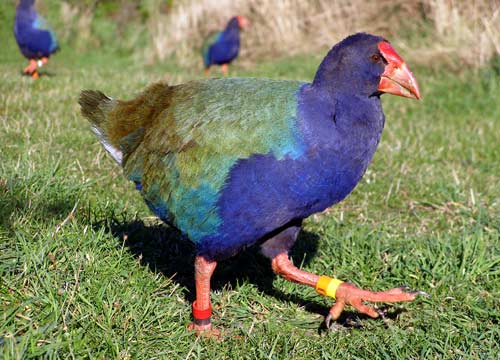
Wildlife medicine in New Zealand is somewhat unique. There are a lot of critters here that don't exist anywhere else, and, conversely, things that aren't here that are everywhere else. During my time on wildbase, I sadly never got to see a snake or ferret. But I did get to see lots of super endangered native creatures, such as takahe. There's like 260 of them left.
And tuatara, which are basically dinosaurs, and the star of today's blog post.
They're basically prehistoric. They're so primitive, they're their own order. I know they look like lizards, but they're closer to dinosaurs. According to wikipedia, "The two species of tuatara are the only surviving members of their order, which flourished around 200 million years ago."
One of them came to the hospital with a diseased eye. It had probably been traumatised (eg poked it into a stick or something) and was now blind, or at least that's my understanding. All their heroic efforts to treat it medically had failed, so it came down to surgery. The first eye enucleation surgery to ever be performed in a tuatara.
The interesting thing about wildlife and exotic medicine is that there isn't a whole mountain of literature for every single species. You might be the second person ever to anaesthetise a giraffe with that combination of drugs, so all you have to go on is one twenty-year old case report and extrapolation from other species. Unfortunately that's not as easy as it sounds, because drugs can have dramatically different effects as species have small differences in enzymes or receptors or whatever. For instance, cats are massively different to dogs in some areas, and we have lots of research about those species. You can imagine how often we diagnose and treat the same disease in, say, a tiger. Now imagine how likely you are to find anything published about a species that numbers in the thousands.
The other fun thing about tuatara surgery is that reptiles are ridiculous animals. The way it was explained to me is that everything in birds happens super fast, they can turn on a dime, and everything that happens in reptiles happens super slow. If a reptile emergency comes in, the first thing you should do is put on the kettle, so you can have a think about it over your tea. Any changes in their health tend to take a long time to manifest. That is important information for this story.
We went ahead with the surgery, which was all quite exciting. It was especially exciting for anaesthesia, who were very stressed out. I don't remember the details because I wasn't on anaesthesia then, but things like heart rate and respiratory rate were apparently quite distressing. So while anaesthesia was freaking out, the wildlife clinician was calmly shrugging it off, with the explanation that some crocodillians can have heart rates as low as one beat per minute. Turtle hearts can keep beating for 24 hours after death (as in, when you open them up at necropsy, you can find their heart beating in front of you... even if you take it out, apparently). Basically, it's impossible to tell if they're dead or alive while under anaesthesia, so he decided not to worry about it. (It fits the theme of not being able to tell if animals are alive while on wildlife roster).
The surgery went along just fine. They packed the empty eye socket with some mini absorbable sponge thingies and sutured it all up. They made it a little bandage that made it look like a pirate. I have an adorable picture, but I doubt I'm supposed to share it on the internet (it might get picked up by google images or something and be stuck there forever when people search about tuatara, and then I think the university might get a teeny bit mad at me).
Details aside, things were fine and dandy that night and into the next morning. During the day, however, the tuatara slowed down, and in grand reptile fashion, we began to question whether it was alive or not. The clinicians broke out more heroics and it went under intensive care for most of the afternoon. Interestingly, since tuatara are in their own order, you need another tuatara if you want to do a blood transfusion... and there's not too many of them around, as you can imagine.
We had the little guy on oxygen, breathing for him, and all sorts of monitoring. He got drugs and fluids and blood products and heating pads and everything we could think of. Unfortunately the monitoring equipment isn't exactly built for tuatara, so the accuracy was questionable, making it even harder to tell anything about the heart or whatever. The heart rate went down and he wouldn't breathe for himself. We did what we could, and gave it a few hours for good measure. He had gone very pale. Surprisingly, we still couldn't figure out if he was actually dead, so we stopped breathing for him and let the chips fall as they may. We put him back in the incubator for the night, just to be sure. The next morinng he hadn't moved any, so we were getting pretty confident we'd lost him.
Too bad for the world's first enucleation on a tuatara. The upside of taking two days to die is that we'd all kind of... accepted it. By the time we gave up on him, I had plenty of time to prepare myself emotionally so I wasn't particularly upset. If he'd given up the ghost in the middle of surgery or something, that would have been more difficult to deal with.
And tuatara, which are basically dinosaurs, and the star of today's blog post.
They're basically prehistoric. They're so primitive, they're their own order. I know they look like lizards, but they're closer to dinosaurs. According to wikipedia, "The two species of tuatara are the only surviving members of their order, which flourished around 200 million years ago."
One of them came to the hospital with a diseased eye. It had probably been traumatised (eg poked it into a stick or something) and was now blind, or at least that's my understanding. All their heroic efforts to treat it medically had failed, so it came down to surgery. The first eye enucleation surgery to ever be performed in a tuatara.
The interesting thing about wildlife and exotic medicine is that there isn't a whole mountain of literature for every single species. You might be the second person ever to anaesthetise a giraffe with that combination of drugs, so all you have to go on is one twenty-year old case report and extrapolation from other species. Unfortunately that's not as easy as it sounds, because drugs can have dramatically different effects as species have small differences in enzymes or receptors or whatever. For instance, cats are massively different to dogs in some areas, and we have lots of research about those species. You can imagine how often we diagnose and treat the same disease in, say, a tiger. Now imagine how likely you are to find anything published about a species that numbers in the thousands.
The other fun thing about tuatara surgery is that reptiles are ridiculous animals. The way it was explained to me is that everything in birds happens super fast, they can turn on a dime, and everything that happens in reptiles happens super slow. If a reptile emergency comes in, the first thing you should do is put on the kettle, so you can have a think about it over your tea. Any changes in their health tend to take a long time to manifest. That is important information for this story.
We went ahead with the surgery, which was all quite exciting. It was especially exciting for anaesthesia, who were very stressed out. I don't remember the details because I wasn't on anaesthesia then, but things like heart rate and respiratory rate were apparently quite distressing. So while anaesthesia was freaking out, the wildlife clinician was calmly shrugging it off, with the explanation that some crocodillians can have heart rates as low as one beat per minute. Turtle hearts can keep beating for 24 hours after death (as in, when you open them up at necropsy, you can find their heart beating in front of you... even if you take it out, apparently). Basically, it's impossible to tell if they're dead or alive while under anaesthesia, so he decided not to worry about it. (It fits the theme of not being able to tell if animals are alive while on wildlife roster).
The surgery went along just fine. They packed the empty eye socket with some mini absorbable sponge thingies and sutured it all up. They made it a little bandage that made it look like a pirate. I have an adorable picture, but I doubt I'm supposed to share it on the internet (it might get picked up by google images or something and be stuck there forever when people search about tuatara, and then I think the university might get a teeny bit mad at me).
Details aside, things were fine and dandy that night and into the next morning. During the day, however, the tuatara slowed down, and in grand reptile fashion, we began to question whether it was alive or not. The clinicians broke out more heroics and it went under intensive care for most of the afternoon. Interestingly, since tuatara are in their own order, you need another tuatara if you want to do a blood transfusion... and there's not too many of them around, as you can imagine.
We had the little guy on oxygen, breathing for him, and all sorts of monitoring. He got drugs and fluids and blood products and heating pads and everything we could think of. Unfortunately the monitoring equipment isn't exactly built for tuatara, so the accuracy was questionable, making it even harder to tell anything about the heart or whatever. The heart rate went down and he wouldn't breathe for himself. We did what we could, and gave it a few hours for good measure. He had gone very pale. Surprisingly, we still couldn't figure out if he was actually dead, so we stopped breathing for him and let the chips fall as they may. We put him back in the incubator for the night, just to be sure. The next morinng he hadn't moved any, so we were getting pretty confident we'd lost him.
Too bad for the world's first enucleation on a tuatara. The upside of taking two days to die is that we'd all kind of... accepted it. By the time we gave up on him, I had plenty of time to prepare myself emotionally so I wasn't particularly upset. If he'd given up the ghost in the middle of surgery or something, that would have been more difficult to deal with.
Friday, 18 July 2014
I Promise I Work in a Real Hospital
A sick rabbit came in today for not eating. We took some radiographs and figured out that it had intestines full of hair balls (trichobezoars). It got sent home with instructions to feed it pineapple juice.
PS: This may be an oversimplification for dramatic effect. We gave it metoclopramide and critical care diet mix, too.
PPS: The reason this is tagged for "neat facts" is that this is a common problem in rabbits and pineapple juice is a commonly suggested aspect of treatment. Apparently it's best if you blend the whole pineapple, since there's a lot of fibre in the core.
PS: This may be an oversimplification for dramatic effect. We gave it metoclopramide and critical care diet mix, too.
PPS: The reason this is tagged for "neat facts" is that this is a common problem in rabbits and pineapple juice is a commonly suggested aspect of treatment. Apparently it's best if you blend the whole pineapple, since there's a lot of fibre in the core.
Saturday, 8 February 2014
The Mysterious Myasthenic, and Acupuncture on Dogs
My week on referral medicine turned out to be as advertised: busy, complicated patients, extra reading, and lots of cool procedures. I alternated between "I am so gonna specialise in internal medicine" and "OMG I just want to be a GP this is too stressful."
Since other groups have purportedly had difficulties with people hogging cases and whatnot, my group learned from their mistakes and picked names randomly to form the order that we would take cases. My name got drawn first, so I took the patient that came in on monday: a four-year-old dog, J. The notes said regurgitation and evidence of megaoesophagus on radiographs - cool, I find oesophageal disease very interesting.
He did not come in with a presentation of oesophageal disease. He was, in fact, making a horrible inspiratory respiratory noise that sounded pretty much exactly like laryngeal paralysis. For some reason, the owner wasn't particularly concerned about this fact, and was a lot more worried about the ongoing "vomiting" the dog had been having since his stay in the kennel. This also happened to be my observed consult which made the whole scenario extra awkward. I proceeded with the consult as normal, but needless to say, we got the dog into the back and on oxygen pretty quickly.
So day 1 was all about stabilising him and figuring out what was going on. We got him into the O2 cage, got a catheter in (with difficulty), and gave him puffs from an inhaler just like people use. We planned to transfer him to surgery, and got him down to anaesthesia to have a laryngeal exam under sedation before they went ahead with the treatment (a laryngeal tie-back).
... He didn't have laryngeal paralysis. His larynx was fine.
Time for a new diagnosis, doctor. It's not like there are a thousand options on the differential diagnosis list... Also, we repeated the radiographs, and did indeed find megaoesophagus. Could it really be myasthenia gravis, the autoimmune disease that attacks the neuromuscular junction?
So J went for a neuro exam, with the boarded veterinary neurologist. She is very good at what she does. She can break down this really complicated subject into something very understandable and her methodical, logical approach to a diagnosis is mind-blowing. Unfortunately, the neuro exam ended up with a really long list of bizarre and subtle symptoms, like short steps, droopy eyes, uneven pupils, and difficulty swallowing. We went over every possible nerve involved and decided it must be a neuropathy or myopathy of some kind, and dysautonomia was a possibility (screwy autonomic nervous system). Putting everything together, myasthenia gravis was still at the top of the list.
There is a really cool test you can do for MG, where you inject a drug and, if they have the disease, they magically get better. Like, instantly. It wears off after a few minutes, but then you know they just need the longer-acting form of that drug and they're good to go. The downside is that an overdose of the drug causes a spectacular crisis. So we tried this--cautiously--even though J's presentation was really weird. And guess what! They decided that he did, indeed, improve--his gait got better and his swallowing improved. Thus he got put onto neostygmine.
All seemed to be going well. Then, when I arrived the next morning, it turns out he ended up having a cholinergic crisis a few hours prior--the whole works: excitement, urination, defaecation, salivation, and all that. Very spectacular and stressful, especially because he pulled out his IV catheter and that made it hard to get the emergency drugs into him. So we fiddled with his dose, and he kept having crises. We switched him from IV drugs to oral drugs (they're absorbed more slwoly), and he kept having crises. Long story short, he didn't respond well to the drugs. His disease symptoms would improve dramatically, but a few hours after the meds he would just have another crisis. He also kept regurgitating, more and more as time went on, and after a while it become yellow, opaque, pus-like gunk. Not good.
At one point, we wanted to use fluoroscopy to identify the best consistency of food for his megaoesophagus. Feeding is a big ordeal because you don't want them to regurgitate it immediately and then inhale it, leading to aspitation pneumonia. The radiology crew got all excited, brought out the fluoroscope, lots of vets, a mob of students, everyone in their lead-lined gowns and all the imaging equipment set up... and J was very stressed out. He got stressed out even with small amounts of handling, so between so many people, and having to put him up onto the imaging table, he went into considerable respiratory distress again. So... "Sorry guys." Everyone dis-gowned and filed out. The radiology crew were very much "Oh no it's perfectly okay!" but I think the students were disappointed, because the fluoroscope isn't used often (it's like real-time x-ray).
The neurologist suggested we try electroacupuncture. It doesn't perform miracles, but it's an effective adjunctive treatment with proven science. And it's awesome. She had just got the gadget so it was her second EAP ever (she does normal acupuncture as well, it's just the electro part that was new, I believe). Using anatomical landmarks such as counting vertebrae, she placed these very very tiny needles into the skin, and connected them to lightweight wires. Even the needles alone were enough to cause endorphin release, and our agitated, nervous dog zonked out completely. When she turned on the current, it was total nap-time for him. It was performed in a quiet consult room.
The way the EAP works is basically using a reflex arc. It stimulates the dermatomes that connect to the spinal cord segments of interest. The signals go in, excite the spinal cord, and in turn signals go out to other muscles. In this case, it was to the oesophagus. The increased electrical activity stimulates the muscles to contract, which they aren't doing normally because of the disease. Then, hopefully, the contraction stimulated by EAP allows the body to regain some of its own, natural tone. She had no idea how effective it would be. However, in her previous patient, an arthritic dog, the EAP proved to be an amazing analgesic, and that dog was able to get up and run and jump almost like normal for a while after the EAP had been performed.
On the thursday, poor J was still having all those troubles with his meds. He'd had 2 sessions of EAP, we'd fiddled with his dose a ton, and he'd still been having regular crises. Then we noticed he was coughing and regurgitating purulent material, took more radiographs, and discovered what we had feared all week: he had aspirated and now had pneumonia. It's possible it had been brewing before he even arrived. It's possible he aspirated some of his food or water while in the hospital, since it's so difficult to manage a patient with megaoesopahgus, and he'd been regurgitating so much. But either way, it became a serious, intensive care situation. Back into the O2 cage, regular monitoring, three or four different IV antibiotics, and a plethora of nursing care requirements. That day was a holiday and I was only scheduled to come in for morning treatments (usually an hour at most), but I ended up there for 5 hours, between taking xrays, writing up the new patient management sheet/requirements, drawing up drugs, monitoring, and administering everything.
This would quickly turn into an extremely expensive, long-term ordeal. We were fairly confident we could get him through this bout of pneumonia, but the problem was that he was very likely to simply aspirate again. The megaoesophagus was likely to be a lifelong management issue, and he was not tolerating his drugs even at low doses. So on the friday, the owners elected to euthanise him. He has been sent to post-mortem to confirm our diagnosis, but I don't know the results of that. It was very sad because he had a dedicated owner, and we had all worked so hard.
Because it was such an interesting, unique, and complex case, this will be my presentation for Grand Rounds this year. There was a great deal of learning for a great deal of students, I got the opportunity to be hugely involved in the patient care, and it was a rare opportunity to see the tests and procedures involved with this rare disease. All in all, it was a great case to have, and I think my week on referral medicine gave a great taste of the many facets of internal medicine.
Since other groups have purportedly had difficulties with people hogging cases and whatnot, my group learned from their mistakes and picked names randomly to form the order that we would take cases. My name got drawn first, so I took the patient that came in on monday: a four-year-old dog, J. The notes said regurgitation and evidence of megaoesophagus on radiographs - cool, I find oesophageal disease very interesting.
He did not come in with a presentation of oesophageal disease. He was, in fact, making a horrible inspiratory respiratory noise that sounded pretty much exactly like laryngeal paralysis. For some reason, the owner wasn't particularly concerned about this fact, and was a lot more worried about the ongoing "vomiting" the dog had been having since his stay in the kennel. This also happened to be my observed consult which made the whole scenario extra awkward. I proceeded with the consult as normal, but needless to say, we got the dog into the back and on oxygen pretty quickly.
So day 1 was all about stabilising him and figuring out what was going on. We got him into the O2 cage, got a catheter in (with difficulty), and gave him puffs from an inhaler just like people use. We planned to transfer him to surgery, and got him down to anaesthesia to have a laryngeal exam under sedation before they went ahead with the treatment (a laryngeal tie-back).
... He didn't have laryngeal paralysis. His larynx was fine.
Time for a new diagnosis, doctor. It's not like there are a thousand options on the differential diagnosis list... Also, we repeated the radiographs, and did indeed find megaoesophagus. Could it really be myasthenia gravis, the autoimmune disease that attacks the neuromuscular junction?
So J went for a neuro exam, with the boarded veterinary neurologist. She is very good at what she does. She can break down this really complicated subject into something very understandable and her methodical, logical approach to a diagnosis is mind-blowing. Unfortunately, the neuro exam ended up with a really long list of bizarre and subtle symptoms, like short steps, droopy eyes, uneven pupils, and difficulty swallowing. We went over every possible nerve involved and decided it must be a neuropathy or myopathy of some kind, and dysautonomia was a possibility (screwy autonomic nervous system). Putting everything together, myasthenia gravis was still at the top of the list.
There is a really cool test you can do for MG, where you inject a drug and, if they have the disease, they magically get better. Like, instantly. It wears off after a few minutes, but then you know they just need the longer-acting form of that drug and they're good to go. The downside is that an overdose of the drug causes a spectacular crisis. So we tried this--cautiously--even though J's presentation was really weird. And guess what! They decided that he did, indeed, improve--his gait got better and his swallowing improved. Thus he got put onto neostygmine.
All seemed to be going well. Then, when I arrived the next morning, it turns out he ended up having a cholinergic crisis a few hours prior--the whole works: excitement, urination, defaecation, salivation, and all that. Very spectacular and stressful, especially because he pulled out his IV catheter and that made it hard to get the emergency drugs into him. So we fiddled with his dose, and he kept having crises. We switched him from IV drugs to oral drugs (they're absorbed more slwoly), and he kept having crises. Long story short, he didn't respond well to the drugs. His disease symptoms would improve dramatically, but a few hours after the meds he would just have another crisis. He also kept regurgitating, more and more as time went on, and after a while it become yellow, opaque, pus-like gunk. Not good.
At one point, we wanted to use fluoroscopy to identify the best consistency of food for his megaoesophagus. Feeding is a big ordeal because you don't want them to regurgitate it immediately and then inhale it, leading to aspitation pneumonia. The radiology crew got all excited, brought out the fluoroscope, lots of vets, a mob of students, everyone in their lead-lined gowns and all the imaging equipment set up... and J was very stressed out. He got stressed out even with small amounts of handling, so between so many people, and having to put him up onto the imaging table, he went into considerable respiratory distress again. So... "Sorry guys." Everyone dis-gowned and filed out. The radiology crew were very much "Oh no it's perfectly okay!" but I think the students were disappointed, because the fluoroscope isn't used often (it's like real-time x-ray).
The neurologist suggested we try electroacupuncture. It doesn't perform miracles, but it's an effective adjunctive treatment with proven science. And it's awesome. She had just got the gadget so it was her second EAP ever (she does normal acupuncture as well, it's just the electro part that was new, I believe). Using anatomical landmarks such as counting vertebrae, she placed these very very tiny needles into the skin, and connected them to lightweight wires. Even the needles alone were enough to cause endorphin release, and our agitated, nervous dog zonked out completely. When she turned on the current, it was total nap-time for him. It was performed in a quiet consult room.
The way the EAP works is basically using a reflex arc. It stimulates the dermatomes that connect to the spinal cord segments of interest. The signals go in, excite the spinal cord, and in turn signals go out to other muscles. In this case, it was to the oesophagus. The increased electrical activity stimulates the muscles to contract, which they aren't doing normally because of the disease. Then, hopefully, the contraction stimulated by EAP allows the body to regain some of its own, natural tone. She had no idea how effective it would be. However, in her previous patient, an arthritic dog, the EAP proved to be an amazing analgesic, and that dog was able to get up and run and jump almost like normal for a while after the EAP had been performed.
On the thursday, poor J was still having all those troubles with his meds. He'd had 2 sessions of EAP, we'd fiddled with his dose a ton, and he'd still been having regular crises. Then we noticed he was coughing and regurgitating purulent material, took more radiographs, and discovered what we had feared all week: he had aspirated and now had pneumonia. It's possible it had been brewing before he even arrived. It's possible he aspirated some of his food or water while in the hospital, since it's so difficult to manage a patient with megaoesopahgus, and he'd been regurgitating so much. But either way, it became a serious, intensive care situation. Back into the O2 cage, regular monitoring, three or four different IV antibiotics, and a plethora of nursing care requirements. That day was a holiday and I was only scheduled to come in for morning treatments (usually an hour at most), but I ended up there for 5 hours, between taking xrays, writing up the new patient management sheet/requirements, drawing up drugs, monitoring, and administering everything.
This would quickly turn into an extremely expensive, long-term ordeal. We were fairly confident we could get him through this bout of pneumonia, but the problem was that he was very likely to simply aspirate again. The megaoesophagus was likely to be a lifelong management issue, and he was not tolerating his drugs even at low doses. So on the friday, the owners elected to euthanise him. He has been sent to post-mortem to confirm our diagnosis, but I don't know the results of that. It was very sad because he had a dedicated owner, and we had all worked so hard.
Because it was such an interesting, unique, and complex case, this will be my presentation for Grand Rounds this year. There was a great deal of learning for a great deal of students, I got the opportunity to be hugely involved in the patient care, and it was a rare opportunity to see the tests and procedures involved with this rare disease. All in all, it was a great case to have, and I think my week on referral medicine gave a great taste of the many facets of internal medicine.
Monday, 24 June 2013
Professors: Viruses
Our virology professor was this sweet, tall, Polish woman. I probably liked her lectures 10x more just because of her accent.
My classmates didn't really like virology, because it was basically learning a long list of viruses. We went through a bunch of families, learning a bunch of details about one virus after another. It was also in this horrible room where the desk is slanted and there's not much space between it and the seats. This means that not only do all your pens and papers slide off the desk, you can't really bend down to get it. If you try, the only option is basically planting your face in your neighbour's lap.
The best part about virology was that she regularly showed us videos in class. I say it's the best part, because it was interesting and a good way to learn, but it was also terrifying. Early on, when we were doing the more routine viruses, they were more simple videos about diagnosis and transmission etc. Then, as we got to the horrible, deadly, zoonotic viruses, we got to see documentaries about how scary and bad such viruses are. For instance there was an emerging virus in Australia that killed some horses... and then the vet who did the post mortem. And the horse's trainer. This was a brand new virus that no one knew about, and now they've done a lot of science and figured out it's in the same family as other wonderful diseases such as canine distemper and rinderpest. Then there was that virus's friend, in pigs in Malasia, that everyone thought was something else transmitted by mosquitoes. So they gave all the pig workers lots of mosquito protection, but a bunch of people died anyway, and they had to quarantine the pig farms and destroy the pigs. Both of those viruses are actually transmitted by fruit bats, particularly their faeces.
Then, of course, there are the fun videos about rabies, documenting not only rabid animals, but rabid people, including interviewing the one girl who is the only person ever to survive it. It kills a significant number of people every year, largely in Africa I believe. That girl had been bitten by a bat (I think she lived in the US), and didn't think to take any precautions. When she came down with the symptoms later, they went through great heroics, and eventually saved her by inducing a coma and keeping the virus from replicating in her nervous system.
Somehow, these things didn't seem to phase our professor. She was as excited about viruses as I think people can get. When she showed us a frightening video about ebola, she was saying how that sort of crazy, big-deal virus that everyone hears about was what got her interested in the discipline initially. You know, the ones that kill practically everybody that catches them. I agree that they are fascinating but I don't think I would be able to sleep at night if I studied them.
I think the thing I learned most from that class was never, ever touch a bat. And it is probably also a good idea to never, ever visit Africa either.
My classmates didn't really like virology, because it was basically learning a long list of viruses. We went through a bunch of families, learning a bunch of details about one virus after another. It was also in this horrible room where the desk is slanted and there's not much space between it and the seats. This means that not only do all your pens and papers slide off the desk, you can't really bend down to get it. If you try, the only option is basically planting your face in your neighbour's lap.
The best part about virology was that she regularly showed us videos in class. I say it's the best part, because it was interesting and a good way to learn, but it was also terrifying. Early on, when we were doing the more routine viruses, they were more simple videos about diagnosis and transmission etc. Then, as we got to the horrible, deadly, zoonotic viruses, we got to see documentaries about how scary and bad such viruses are. For instance there was an emerging virus in Australia that killed some horses... and then the vet who did the post mortem. And the horse's trainer. This was a brand new virus that no one knew about, and now they've done a lot of science and figured out it's in the same family as other wonderful diseases such as canine distemper and rinderpest. Then there was that virus's friend, in pigs in Malasia, that everyone thought was something else transmitted by mosquitoes. So they gave all the pig workers lots of mosquito protection, but a bunch of people died anyway, and they had to quarantine the pig farms and destroy the pigs. Both of those viruses are actually transmitted by fruit bats, particularly their faeces.
Then, of course, there are the fun videos about rabies, documenting not only rabid animals, but rabid people, including interviewing the one girl who is the only person ever to survive it. It kills a significant number of people every year, largely in Africa I believe. That girl had been bitten by a bat (I think she lived in the US), and didn't think to take any precautions. When she came down with the symptoms later, they went through great heroics, and eventually saved her by inducing a coma and keeping the virus from replicating in her nervous system.
Somehow, these things didn't seem to phase our professor. She was as excited about viruses as I think people can get. When she showed us a frightening video about ebola, she was saying how that sort of crazy, big-deal virus that everyone hears about was what got her interested in the discipline initially. You know, the ones that kill practically everybody that catches them. I agree that they are fascinating but I don't think I would be able to sleep at night if I studied them.
I think the thing I learned most from that class was never, ever touch a bat. And it is probably also a good idea to never, ever visit Africa either.
Wednesday, 19 June 2013
Try it Five Times Fast
Having recently completed the mid-year exam for small animal medicine and surgery, I thought I would share some of the words that are amazingly fun. I actually understand all these.
Cholecystoduodenostomy
Hypergammaglobulinaemia
Oesophagogastroduodenoscopy
Parasympathomimetic
Pseudohyperparathyroidism
There are, of course, many more where that came from.
I would also like to add, what is not fun is not having a good way to shorten "apocrine gland adenocarcinoma of anal sac origin" and having to write that every time a professor talks about. (Note: I recently learned I could use AGASACA and it made my entire week)
Cholecystoduodenostomy
Hypergammaglobulinaemia
Oesophagogastroduodenoscopy
Parasympathomimetic
Pseudohyperparathyroidism
There are, of course, many more where that came from.
I would also like to add, what is not fun is not having a good way to shorten "apocrine gland adenocarcinoma of anal sac origin" and having to write that every time a professor talks about. (Note: I recently learned I could use AGASACA and it made my entire week)
Friday, 14 June 2013
What Cell Would You Be?
In clin path, when we were learning about eosinophils, our professor mentioned that the pathologists had once had a discussion about what cell you would be, or what cell describes your personality. (Because pathologists are the biggest nerds out of all of us)
She brought it up because she really loves horse eosinophils. They look sort of like raspberries.
There are a lot of cells I like, and some that I'm more partial to than others. My friend and I once talked about this in parasites--there were certain ones that spoke to us so we never got them wrong if they came up on a test. One of mine was Ancylostoma caninum, a hookworm in dogs.
She brought it up because she really loves horse eosinophils. They look sort of like raspberries.
There are a lot of cells I like, and some that I'm more partial to than others. My friend and I once talked about this in parasites--there were certain ones that spoke to us so we never got them wrong if they came up on a test. One of mine was Ancylostoma caninum, a hookworm in dogs.
Another was Toxocara canis because it is the same sort of worm as Dune sandworms.
But back to the topic, there are some nice cells to choose from. I quite like neutrophils because they stand out so clean and nicely on a blood smear.
They can be a little ragged, however. My favourite is one that is much more pristine--very smooth and fine: the spherocyte. Even the name has a nice roundness!
These are cells formed by partial phagocytosis--a macrophage takes a chomp out of a red blood cell, so the RBC stretches its membrane over the defect and gets smaller and more compact. They're present in immune-mediated haemolytic anaemia.
Saturday, 6 October 2012
Dog Hearts
Unlike people, heart attacks are very rare in dogs. Come to think of it, they are rare in all domestic animals.
Interestingly about dogs, they do get high cholesterol commonly. This is because a lot of dogs have hypothyroidism, and it causes high cholesterol (along with several other diseases). However, it doesn't tend to do anything to their coronary vessels and they are just fine.
Thursday, 4 October 2012
A Bit About Pathology
In third year we have a double semester paper on pathology. It starts out as general path, where we learn basics of pathological processes, such as what's going on when there's inflammation or necrosis, and the rest of the year we go system by system learning about the common things that go wrong. You wouldn't believe the things that happen in the body!
Here are some of the ones I thought were interesting, I'll pick one from a few different systems:
Cardiovascular System - Patent Ductus Arteriosus
A fetus doesn't use its lungs, so its whole circulation is set up differently. It gets everything it needs from the placenta, and the lungs just stay uninflated and all the blood bypasses them. At birth, when the neonate takes its first breath and inflates the lungs, all the blood rushes to them and the bypasses shut off. There's a number of congenital diseases where those fetal shunts don't close properly.
For instance, there's a channel called the ductus arteriosus which, in the fetus, allows blood that would have gone to the lungs just go straight into the aorta instead and off to the rest of the body. Sometimes it doesn't close in the newborn. The interesting thing is that now, there's space in the lungs to fill, so rather than blood hopping into the aorta, it flows preferentially back out of the aorta and to the lungs (how much blood depends on how big the hole is). So you get blood that circulates uselessly between the heart and the lungs without ever going to the body.
Endocrine System - Pituitary Cyst
Honestly, the endocrine system is really cool, and I could put down any one of the diseases we learned about and be really excited, but they also all require long-winded explanations of what the hormones do and why you see the pathology that you do. So I thought I would show you a picture of a dog with a pituitary cyst, causing a lack of pituitary hormones including growth hormones (a pituitary dwarf). These dogs are littermates:
Gastrointestinal System - Rumen Acidosis
If you feed a cow too much grain, you can end up with this terrible domino effect of consequences. If they aren't used to eating grain and get a whole bunch at once, it screws up the microbes in the flora, and they start fermenting all the carbohydrate to produce acid. The lowered pH is called acidosis, and causes inflammation in the rumen wall, mucking up absorption of nutrients. This inflammation makes the wall weak and necrotic, so bacteria and fungi have a party, even making it into the blood.
Blood from the GI tract goes to the liver for processing, so the bacteria hitch a ride and hop out, causing liver abscesses. This causes clots to form in the big veins heading out of the liver back to the heart. The next place the blood goes is the lungs, so the clots can break off and float out there, and then get stuck in the lungs. This causes an aneurysm (outpouching of the blood vessel) which eventually pops and all the blood flows out into the airways.
The cow proceeds to die a very dramatic and rapid death with blood pouring profusely from its nostrils. All because it ate a bunch of grain a while back.
Genital System - Teratomas
If you think about it, cells in the ovary are about as undifferentiated as you can get: eggs go on to create a new individual and divide into cells that form every tissue in the body. So if one of them goes out of control, it's not like a skin cell that does skin things or a liver cell that does liver things; its an embryo cell that does embryo things. So these tumours have all kinds of crazy things in them, like fully formed teeth, hairs, and glands.
Liver - Hepatogenous Photosensitivity
For animals that eat plants, like cows, microbes in the GI tract break down all the chlorophyll they ingest, and this forms a compound called phylloerythrin that gets absorbed into the blood. It's the liver's job to take care of that stuff and get rid of it. If the liver gets munted, the phylloerythrin builds up.
You know how chlorophyll absorbs sunlight? This breakdown product floating around in the blood also reacts to sunlight. Once it makes it to the skin, it absorbs and releases energy from the sun, basically making the animal get bad sunburns.
For animals that eat plants, like cows, microbes in the GI tract break down all the chlorophyll they ingest, and this forms a compound called phylloerythrin that gets absorbed into the blood. It's the liver's job to take care of that stuff and get rid of it. If the liver gets munted, the phylloerythrin builds up.
You know how chlorophyll absorbs sunlight? This breakdown product floating around in the blood also reacts to sunlight. Once it makes it to the skin, it absorbs and releases energy from the sun, basically making the animal get bad sunburns.
Lymphohaemopoeitic System - Hemangiosarcoma
The spleen is involved in monitoring and storing blood, and in dogs it's the main predilection site for a type of cancer called a hemangiosarcoma. This is basically cancer of the blood vessels, and I think it's really interesting because, since it's the cells that make up the vessel walls, when they proliferate they try to form new blood vessels. Since they're cancer cells, they really suck at it, so the result is a bunch of shitty weak random vessels that are prone to rupture. Unfortunately this type of cancer is very deadly (it's most common in dogs).
Skeletal Muscle - Myotonia
This is actually a terrible and tragic genetic disease in people, horses, dogs, and cats, but for some reason goats have a perfectly harmless version.
Monday, 1 October 2012
Brachycephalic Airway Syndrome
They live their entire lives snorting and snuffling because they have tissue falling down the back of their throat. When we anaesthetise dogs we put a tube down their trachea, and most dogs rather resent it when they're waking up. These dogs actually don't mind because it's probably the first time in their lives they can actually breathe.
Brachycephalic airway syndrome is a combination of stenotic (narrow) nostrils and an excessively long soft palate. That is the tissue that separates your nose from your mouth near the back. The theory is that as we breed for shorter faces, the genes for bones respond and the skull gets short, but the soft tissue doesn't respond as quickly, so that's relatively too big. What's even worse is that bulldogs commonly have hypoplastic (underdeveloped) tracheas, so not only is there a bunch of tissue to try and breathe around, they are also breathing through a straw.
So remember, if you buy one of these dogs, you are supporting the breeding of dogs with a genetic disease. You may think its cute but your dog will live its entire life without being able to breathe properly. I don't think it's very fair to the animal.
Sunday, 30 September 2012
There's Something Fishy About This Lab
A week or two ago, we had a fish handling lab.
Did you know that people anaesthetise fish? Fish are sedated before they are killed commercially, but people also have valuable pet fish or display fish (like in an aquarium) that sometimes get sick and need veterinary care. There are a number of basic diagnostic techniques for investigating what could be wrong with a sick fish, and medicine or even surgery can be used to treat them.
Fish anaesthesia is surprisingly like mammalian anaesthesia. For a dog, the anaesthetic is vaporised and they breathe it in through their lungs via the anaesthetic machine. For a fish, it's basically the same: the drug is dissolved in the water rather than the air, and they "breathe" it in through their gills. If a dog is too deep, you give it pure oxygen to breathe for a while; for a fish, you put them into fresh water to breathe for a while.
Unfortunately for me, I managed to pick crappy fish for my partner and myself. They would not go to sleep. As they get more sedated, they lose their balance reflexes (float sideways and stuff), but it can freak them out a little as they go down. So one would have a panic attack, and that would wake up the other one, which was just drifting off. Then the first one would calm down, but the second one got a fright. The professor came by to increase the dose three times after the first lot! We spent at least half of our lab time staring into the bucket waiting for them to be deep enough to work with. It didn't help that we kept thinking they were fine, picking them up, and then they would flop around and wake up and we'd have to put them back. As an aside, the anaesthetic we were using is derived from clove oil, so the lab smelled like a strange combination of live fish and cloves.
Once they were asleep, we could take them out and do things to them. They survive in the air because their metabolism and respiration is slowed down so much under anaesthesia. Normally you have to keep them really wet to protect their skin, however. Our lab was designed to be a non-recovery lab, using fish at the end of their lives and such, so we didn't worry about that.
First thing we did was take blood. There's a vein in their tail, right in front of the fin, and we spent almost the entire rest of our lab time trying to stab it. If the needle goes through a scale on the way in, you plug up the thing with keratin and have to get a new needle. Little fish have little veins and not a lot of blood, so it was a pretty frustrating endeavour. The other site I tried was the dorsal aorta, which sounds really scary: you stick the needle into the back of their mouths, and that's apparently right where the aorta comes off the heart. I blindly stuck mine in and actually got an entire drop of blood right off the bat, but everyone else I talked to had a much harder time with that one.
Another thing we did was skin scrapes, which is apparently quite useful as fish can get protozoal infections, and if you put the mucus on a slide, you can see the protozoa move around. Our fish were not so interesting.
The last thing we did with the live fish was a gill biopsy. If you don't know anything about fish physiology, I have to tell you, gills are cool. They look cool, they are designed cool, they are just cool. You can also snip off a tiny piece with scissors, put it on a slide, and have a look for disease or degeneration that might be making the fish sick (it would be like people having a lung problem). Electron micrograph photo:
The last thing we did was euthanise the fish and do a post mortem. My partner and I had basically no time to do this, so we only had a quick look before lab ended. They have inflated swim bladders next to the kidney, that was the neatest thing, like a little balloon inside their body cavity. They have teeny tiny little spleens, massive massive ovaries filled with eggs, a simple two chambered heart, and skinny twisty intestines. From google:
Did you know that people anaesthetise fish? Fish are sedated before they are killed commercially, but people also have valuable pet fish or display fish (like in an aquarium) that sometimes get sick and need veterinary care. There are a number of basic diagnostic techniques for investigating what could be wrong with a sick fish, and medicine or even surgery can be used to treat them.
Fish anaesthesia is surprisingly like mammalian anaesthesia. For a dog, the anaesthetic is vaporised and they breathe it in through their lungs via the anaesthetic machine. For a fish, it's basically the same: the drug is dissolved in the water rather than the air, and they "breathe" it in through their gills. If a dog is too deep, you give it pure oxygen to breathe for a while; for a fish, you put them into fresh water to breathe for a while.
Unfortunately for me, I managed to pick crappy fish for my partner and myself. They would not go to sleep. As they get more sedated, they lose their balance reflexes (float sideways and stuff), but it can freak them out a little as they go down. So one would have a panic attack, and that would wake up the other one, which was just drifting off. Then the first one would calm down, but the second one got a fright. The professor came by to increase the dose three times after the first lot! We spent at least half of our lab time staring into the bucket waiting for them to be deep enough to work with. It didn't help that we kept thinking they were fine, picking them up, and then they would flop around and wake up and we'd have to put them back. As an aside, the anaesthetic we were using is derived from clove oil, so the lab smelled like a strange combination of live fish and cloves.
Once they were asleep, we could take them out and do things to them. They survive in the air because their metabolism and respiration is slowed down so much under anaesthesia. Normally you have to keep them really wet to protect their skin, however. Our lab was designed to be a non-recovery lab, using fish at the end of their lives and such, so we didn't worry about that.
First thing we did was take blood. There's a vein in their tail, right in front of the fin, and we spent almost the entire rest of our lab time trying to stab it. If the needle goes through a scale on the way in, you plug up the thing with keratin and have to get a new needle. Little fish have little veins and not a lot of blood, so it was a pretty frustrating endeavour. The other site I tried was the dorsal aorta, which sounds really scary: you stick the needle into the back of their mouths, and that's apparently right where the aorta comes off the heart. I blindly stuck mine in and actually got an entire drop of blood right off the bat, but everyone else I talked to had a much harder time with that one.
Another thing we did was skin scrapes, which is apparently quite useful as fish can get protozoal infections, and if you put the mucus on a slide, you can see the protozoa move around. Our fish were not so interesting.
The last thing we did with the live fish was a gill biopsy. If you don't know anything about fish physiology, I have to tell you, gills are cool. They look cool, they are designed cool, they are just cool. You can also snip off a tiny piece with scissors, put it on a slide, and have a look for disease or degeneration that might be making the fish sick (it would be like people having a lung problem). Electron micrograph photo:
The last thing we did was euthanise the fish and do a post mortem. My partner and I had basically no time to do this, so we only had a quick look before lab ended. They have inflated swim bladders next to the kidney, that was the neatest thing, like a little balloon inside their body cavity. They have teeny tiny little spleens, massive massive ovaries filled with eggs, a simple two chambered heart, and skinny twisty intestines. From google:
Thursday, 20 September 2012
Tongue-is Contortus
Parasitology involves a lot of harrowing memorisation, especially since we have to not only recall parasite names, we get points off if we spell them incorrectly. I apologise for the horrible almost-pun of the post title, it amuses me (inspired by our friend, Haemonchus contortus). Anyway, here are some particularly bizarre names...
Funny sounding:
Bunostomum phlebotomum
How do you pronounce this?:
Ollulanus tricuspis
Ornithostrongylus quadriradiatus
Trichostrongylus axei
Winner of all:
Macracanthorhynchus hirudinaceus
There are many others that I find entertaining, but I realised I was turning this into quite a long list, and no one is likely to be that interested.
Funny sounding:
Bunostomum phlebotomum
Dictyocaulus arnfieldi
Nematodirus
helvetianus
Protostrongylus
rufescens
How do you pronounce this?:
Aelurostrongylus abstrusus
Dipetalonema reconditumOllulanus tricuspis
Ornithostrongylus quadriradiatus
Trichostrongylus axei
Uncinaria
stenocephala
Winner of all:
Macracanthorhynchus hirudinaceus
There are many others that I find entertaining, but I realised I was turning this into quite a long list, and no one is likely to be that interested.
Friday, 12 August 2011
Avian Anatomy
Our last anatomy lab was on avian anatomy, so we dissected chickens. Here is an account of some of the things I thought were interesting.
You'd be surprised how few feathers they actually have. When they're alive they're all poofy, but for dissecting they had us dip them in soapy water so that feathers wouldn't be floating everywhere all lab (I'm serious), and you can really see that there's not as many as you'd think.
Birds have a super massive keel on their bellies, part of their sternum. Imagine your sternum down your chest, but with a big fin. Most of their bones are the same as mammals, but lots of things are fused. Most of their vertebral column is fused, which gives them stability and allows them to have less back muscles, which makes them lighter. The wishbone is their clavicle. Another interesting bony structure is the bone in their tongue--they don't have tongue muscles; the movement is conducted through their tongue bone.
Birds have a different voice organ than mammals, called the syrinx (we use our larynx). Something interesting I read is that songbirds and such that make a lot of complex sounds don't necessarily have a more developed syrinx or associated muscles.
Their brains are tiny, which I'm sure you already knew. What you might have not known is that their eyes are huge, way bigger than they appear because they aren't a sphere. Each eye is about the same size as the brain. Also their skull is kind of thick too, so if you're just looking at a bird and try to picture the size of their brain in their head, it's actually way, way smaller than you'd think just by doing that. They can have pretty super vision, but I don't think their brains do a whole lot else for them.
Birds don't have a diaphragm: they depend on moving their body wall to draw air into their lungs. So if you hold a bird so it can't move it's chest, it won't be able to breathe. They also have a pretty cool system, where air gets drawn into these air sacs and then into the lungs and then into other air sacs before being exhaled, so that the air going into the lungs is always fresh. Being so efficient is why they can fly very high, where the oxygen is low.
Their gastrointestinal, urinary, and reproductive tracts all open into this one pouch and exit the bird through the same hole. They sort of have two stomachs--one that's glandular, and one that's super muscular (the gizzard). That's kind of neat because it actually utilises ingested stones to help grind up food. Their testicles don't descend (they stay inside the abdomen), and sometimes the only way to sex a bird might be using internal imaging. They also don't have a bladder.
If you look at the ovary, you can see a bunch of big red follicles, like the ones that will be tomorrow's egg and the next day's. They're super huge! And if you break one by accident, you get yolk everywhere. In our bird, it had actually had a ruptured part of its reproductive tract, so there was an egg that hadn't got its shell yet, and it was just hanging out inside the bird. It looked just like it would have if you'd cracked one and dropped it in.
The nerve that innervates their hind limbs passes over the kidney, so if they have kidney disease or a tumour or something, it can actually present as lameness.
Perching is a passive thing. When they bend their knees, it tenses the tendons that flex their digits, causing them to grip. It takes no energy for them to sit there gripping a branch, and if you want to un-perch them you have to straighten their legs.
Some birds, such as owls, have asymmetric ears so they can localise sound better. Also, birds have magnetite in their beak and neck muscles, which is an iron rich crystal that responds to the Earth's magnetic field. This gives them both directional and geographical location information.
Those are all the cool facts about birds I can think of for now, I hope you learned something.
You'd be surprised how few feathers they actually have. When they're alive they're all poofy, but for dissecting they had us dip them in soapy water so that feathers wouldn't be floating everywhere all lab (I'm serious), and you can really see that there's not as many as you'd think.
Birds have a super massive keel on their bellies, part of their sternum. Imagine your sternum down your chest, but with a big fin. Most of their bones are the same as mammals, but lots of things are fused. Most of their vertebral column is fused, which gives them stability and allows them to have less back muscles, which makes them lighter. The wishbone is their clavicle. Another interesting bony structure is the bone in their tongue--they don't have tongue muscles; the movement is conducted through their tongue bone.
Birds have a different voice organ than mammals, called the syrinx (we use our larynx). Something interesting I read is that songbirds and such that make a lot of complex sounds don't necessarily have a more developed syrinx or associated muscles.
Their brains are tiny, which I'm sure you already knew. What you might have not known is that their eyes are huge, way bigger than they appear because they aren't a sphere. Each eye is about the same size as the brain. Also their skull is kind of thick too, so if you're just looking at a bird and try to picture the size of their brain in their head, it's actually way, way smaller than you'd think just by doing that. They can have pretty super vision, but I don't think their brains do a whole lot else for them.
Birds don't have a diaphragm: they depend on moving their body wall to draw air into their lungs. So if you hold a bird so it can't move it's chest, it won't be able to breathe. They also have a pretty cool system, where air gets drawn into these air sacs and then into the lungs and then into other air sacs before being exhaled, so that the air going into the lungs is always fresh. Being so efficient is why they can fly very high, where the oxygen is low.
Their gastrointestinal, urinary, and reproductive tracts all open into this one pouch and exit the bird through the same hole. They sort of have two stomachs--one that's glandular, and one that's super muscular (the gizzard). That's kind of neat because it actually utilises ingested stones to help grind up food. Their testicles don't descend (they stay inside the abdomen), and sometimes the only way to sex a bird might be using internal imaging. They also don't have a bladder.
If you look at the ovary, you can see a bunch of big red follicles, like the ones that will be tomorrow's egg and the next day's. They're super huge! And if you break one by accident, you get yolk everywhere. In our bird, it had actually had a ruptured part of its reproductive tract, so there was an egg that hadn't got its shell yet, and it was just hanging out inside the bird. It looked just like it would have if you'd cracked one and dropped it in.
The nerve that innervates their hind limbs passes over the kidney, so if they have kidney disease or a tumour or something, it can actually present as lameness.
Perching is a passive thing. When they bend their knees, it tenses the tendons that flex their digits, causing them to grip. It takes no energy for them to sit there gripping a branch, and if you want to un-perch them you have to straighten their legs.
Some birds, such as owls, have asymmetric ears so they can localise sound better. Also, birds have magnetite in their beak and neck muscles, which is an iron rich crystal that responds to the Earth's magnetic field. This gives them both directional and geographical location information.
Those are all the cool facts about birds I can think of for now, I hope you learned something.
Tuesday, 17 May 2011
CPR
SCVECCS is the Student Veterinary Emergency and Critical Care Society, and they have lots of interesting seminars and workshops. Recently I went to a CPR lab.
I've taken human CPR courses before, and chances aren't bad that so have you. I remember having the fake chest and practice defibrillator. Well, this wasn't like that. It was in the wet lab, which is a room kind of like a mini-version of the anatomy lab and all sorts of stuff goes on in there. There were two groups, I was in the second, and the first group went more than an hour over time. When we finally went in, they were still trying to finish up, and the first thing I saw was a room full of vet students sticking needles and tubes into the dead sheep that were sprawled everywhere. They were dirty and bloody and the room smelled rather like blood and sheep, as you might expect. I did actually find it pretty unpleasant at first, but as soon as we started doing stuff it improved a lot, since focusing on the techniques precluded focusing on how disturbing a few of the sheep were.
There were four stations.
I've taken human CPR courses before, and chances aren't bad that so have you. I remember having the fake chest and practice defibrillator. Well, this wasn't like that. It was in the wet lab, which is a room kind of like a mini-version of the anatomy lab and all sorts of stuff goes on in there. There were two groups, I was in the second, and the first group went more than an hour over time. When we finally went in, they were still trying to finish up, and the first thing I saw was a room full of vet students sticking needles and tubes into the dead sheep that were sprawled everywhere. They were dirty and bloody and the room smelled rather like blood and sheep, as you might expect. I did actually find it pretty unpleasant at first, but as soon as we started doing stuff it improved a lot, since focusing on the techniques precluded focusing on how disturbing a few of the sheep were.
There were four stations.
- The first one my group did was intubation, which is sticking a tube down the trachea so the patient can breathe. This is supposedly quite difficult in sheep, and the demonstrator took a long time trying to get it in. When it was our turn, one guy started off on that sheep, so I went over to the second sheep. It was really easy and took like a second for me and the person with me. One cool part is the scope that you stick down their throat; it not only holds down the tongue and other structures out of the way, but has a light on the end so you can see.
- Warning possibly disturbing mental image: the sheep for this one were cut along their cheeks like the Joker from the Dark Knight so we could open their mouths better, since they were in rigour mortis. You can imagine why I thought it was a little creepy at first, when one of the first things I saw upon entering the room was bloody dead Joker sheep.
- The next was putting catheters into veins. This is slightly more difficult in dead animals, as they have no blood pressure. Anyway, there are three main sites you can try (jugular, cephalic, and saphenous veins), and we stuck them all. Maybe. It was kind of hard to tell.
- The third involved drawing blood, and instead of sheep they had rubber dog heads. These things are ancient and full of holes because they've been jabbed so many times, and the tubes inside representing the veins are so leaky we couldn't actually get water out like we were supposed to. They also showed us the crash cart and cave us a little description of what was inside.
- The last station was doing actual chest compressions. There were some more dead sheep, but also an awesome stuffed dog. It was black and long-haired, had a zipper up its belly, and looked kind of like cookie monster with teeth. Unlike humans, you have the animals on their side rather than their back. On really small animals, you can even just use your thumb and fingers to squeeze their chest. Also since they're animals, you have to aim for 100-120 beats per minute, so pretty quick.
Despite all the effort and technique involved in CPR, they told us the recovery rate is 5%. That's counting patients that arrest during anaesthesia. If you only count patients that come in (say, hit by a car), it's down to 1%. Makes it seem a little futile.
Subscribe to:
Posts (Atom)