In a recent anatomy lab, the professor announced that we were to remove the cranial half of our dog and put it into the preserved material bin.
In other words, "Cut off the front of the dog so you only have an abdomen and hind legs, and then toss the head part out." Kind of like that magic trick with the saw and the person in a box.
I was under the impression that at the end of the year we would be able to dissect the side that we haven't touched yet, for revision for the final. Apparently that's not the case, and having a bunch of dissected tissue is just a detriment since we'll only be looking at the abdomen now. After all, even if you take pretty good care of it, you can still get drying out and damage, which is not good for anybody, especially the cadavre. At any rate, we now only have half a dog.
Friday, 13 May 2011

Tuesday, 10 May 2011
Things That Are Creepy to Dissect
Warning: this is more graphic than most posts, and I don't recommend reading it if you get creeped out by mental images.
That being said, here are some of the things that I found disconcerting or downright creepy, as well as a few that I was expecting to be worse than they were.
Paw Pad: In the first semester, we navigated our way through the many muscles of the body, and for a long time nothing we did was at all disturbing. I may have even been a little disappointed, because I was expecting anatomy lab to be a bit crazier (it has since met my expectations this year). Even removing the skin didn't feel very weird. It was all good and fun, until we got to the paw. I'm not sure if I can explain why it was so, but there seemed to be unanimous agreement that removing the paw pad was uncomfortable. You might be able to conjure up the same feeling if you look at the nearest furry animal, and picture their thick, callous paw pad alone on a table, cold, attached to some preserved skin.
Eyelid: If you see below, the eye was cool. What was weird was the surrounding skin that was left behind, including the eyelids and lashes. Getting the eye out was super awkward, and since we had to cut everything out, in the end we were left with the ring of skin, which like I said was weird to look at by itself, so I put it back over the socket. That was a bit creepy. Imagine looking into a dog face, with its eyelids open, and an empty socket behind them.
Tongue: Mostly because the dog is dead and preserved, I don't like touching her tongue. We didn't dissect it or anything, though it did come out attached to the larynx (the Adam's Apple area) when we were looking at that. Normally dog tongues are soft and warm and wet and sloppy, but this one was so dry and cold, and rock hard. Not comfortable to touch when you're used to dogs being not preserved.
Lips and Cheeks: In a similar vein, the lips and cheeks are also a bit weird to see and feel. Probably what made them look so unusual was the fact that we had sawed the jaw in half, so a lot of the time we had it folded back or twisted in a highly unnatural manner, showing the inside of the cheeks or twisting the lips in a way that only added to the preserved-cold-dry feel.
Sawing through the face: Sometimes when we sawed through bone, everything was dissected away, but not all the time. In order to look at the inside of the skull in the muzzle, we put the saw to the nose (which, you might notice the theme, is cold and dry and preserved feeling) and just sawed on through the face. Getting the little section of skull off wasn't very creepy except for that first part, going through the nose. Dog noses are attached by cartilage, and if you have a dog you may have noticed how their noses are very wiggly and pliable. They're still wiggly and pliable after the dog is dead.
Nasal concha: After we did that, we could see into the nasal cavity. The way it works is there's a ton of thin, flat bone (called the nasal concha) that's all curly and covered in mucous, giving it lots of surface area. The air goes over that and dust or whatever crap you're breathing gets stuck on the mucous, and the nice clean air keeps going. For some reason our lab guide told us to remove the nasal concha, I think it was so we could see some openings in the bone (like where ducts open, such as the tear ducts). Since it's so thin and brittle, one of my lab partners just took his thumb and ripped it out. And, oh man, did it make such a haunting crunching noise! It is bone, after all, just very thin bone.
Abdominal Fat: If you've ever seen an abdominal surgery, you'll have seen that there's a sheet of fat and blood vessels that covers the intestines, attached at the top like an apron, which the surgeon has to lift away. This is where fat goes on fat people, and it's called the greater omentum. In the live animal, it's quite pink and fleshy. In the preserved dogs, it's very thin, transparent, and lacy. It's a lot like lacy curtains. It's weird.
Stomach: Dogs eat a lot of gross stuff. Digested dog food is one thing, but ours had tons hair (mostly dog hair), and even worse, a bunch of chewed up bone pieces she must have been fed. The inside of her stomach was pretty full. It was grainy or sandy to the touch. It was not cool.
The things that I expected to be worse:
Ribs: As I mentioned previously, the ribs are sort of in the way if you want to look at stuff in the thorax. What we did was to snap the bones at each end, but leave the muscles and everything attached, so it made a flap we could lift up. Snapping the ribs was actually pretty fun; we used bone cutters that are basically giant wire cutters. Since it was the first bone cutting lab, they had forgotten to put out safety goggles, and I tried to convince my lab group to let me do it since I was the only one with glasses. They weren't fooled, however.
Eyes: This is the classic thing you'd expect to be unsettling, especially when you cut them up and look at the goop inside. As it turns out, I didn't find it nearly as creepy as the empty eye socket, possibly because it's complicated enough to keep you interested in what you're seeing without getting grossed out. The lens was pretty cool too, it was a hard, oval little white thing rather like a hard candy.
Heart: To allow us to see blood vessels in the cadavres, they're filled with latex (red for arteries, blue for veins), so this transforms the hearts into very solid, latex-filled lumps. In order to actually understand the interior of the heart, we were given fresh sheep hearts to handle and dissect. This involved sticking lots of fingers in the chambers, and feeling soft flesh instead of the unnaturally dry, hard preserved material. It was a very good way to see what openings were where and was probably one of the best labs we did.
Brain: As you might have gathered from some previous posts, we took the brain out of our dog. Dog brains are pretty small--I look at my dog and feel pity--and they're shaped differently from a human's, because the spinal cord goes straight out the back rather than downwards from the bottom. It was a rubbery, squishy thing, kind of like those stress balls, and I spent a good portion of the lab being the one to hold it. Later on, we used a "brain knife" to cut it into transverse sections, which is like a small ruler with a sharp edge. That was cool because we could see all the structures inside the brain, and put it back together to see how it works in 3D. Some of the brain labs felt very Young Frankenstein.
That being said, here are some of the things that I found disconcerting or downright creepy, as well as a few that I was expecting to be worse than they were.
Paw Pad: In the first semester, we navigated our way through the many muscles of the body, and for a long time nothing we did was at all disturbing. I may have even been a little disappointed, because I was expecting anatomy lab to be a bit crazier (it has since met my expectations this year). Even removing the skin didn't feel very weird. It was all good and fun, until we got to the paw. I'm not sure if I can explain why it was so, but there seemed to be unanimous agreement that removing the paw pad was uncomfortable. You might be able to conjure up the same feeling if you look at the nearest furry animal, and picture their thick, callous paw pad alone on a table, cold, attached to some preserved skin.
Eyelid: If you see below, the eye was cool. What was weird was the surrounding skin that was left behind, including the eyelids and lashes. Getting the eye out was super awkward, and since we had to cut everything out, in the end we were left with the ring of skin, which like I said was weird to look at by itself, so I put it back over the socket. That was a bit creepy. Imagine looking into a dog face, with its eyelids open, and an empty socket behind them.
Tongue: Mostly because the dog is dead and preserved, I don't like touching her tongue. We didn't dissect it or anything, though it did come out attached to the larynx (the Adam's Apple area) when we were looking at that. Normally dog tongues are soft and warm and wet and sloppy, but this one was so dry and cold, and rock hard. Not comfortable to touch when you're used to dogs being not preserved.
Lips and Cheeks: In a similar vein, the lips and cheeks are also a bit weird to see and feel. Probably what made them look so unusual was the fact that we had sawed the jaw in half, so a lot of the time we had it folded back or twisted in a highly unnatural manner, showing the inside of the cheeks or twisting the lips in a way that only added to the preserved-cold-dry feel.
Sawing through the face: Sometimes when we sawed through bone, everything was dissected away, but not all the time. In order to look at the inside of the skull in the muzzle, we put the saw to the nose (which, you might notice the theme, is cold and dry and preserved feeling) and just sawed on through the face. Getting the little section of skull off wasn't very creepy except for that first part, going through the nose. Dog noses are attached by cartilage, and if you have a dog you may have noticed how their noses are very wiggly and pliable. They're still wiggly and pliable after the dog is dead.
Nasal concha: After we did that, we could see into the nasal cavity. The way it works is there's a ton of thin, flat bone (called the nasal concha) that's all curly and covered in mucous, giving it lots of surface area. The air goes over that and dust or whatever crap you're breathing gets stuck on the mucous, and the nice clean air keeps going. For some reason our lab guide told us to remove the nasal concha, I think it was so we could see some openings in the bone (like where ducts open, such as the tear ducts). Since it's so thin and brittle, one of my lab partners just took his thumb and ripped it out. And, oh man, did it make such a haunting crunching noise! It is bone, after all, just very thin bone.
Abdominal Fat: If you've ever seen an abdominal surgery, you'll have seen that there's a sheet of fat and blood vessels that covers the intestines, attached at the top like an apron, which the surgeon has to lift away. This is where fat goes on fat people, and it's called the greater omentum. In the live animal, it's quite pink and fleshy. In the preserved dogs, it's very thin, transparent, and lacy. It's a lot like lacy curtains. It's weird.
Stomach: Dogs eat a lot of gross stuff. Digested dog food is one thing, but ours had tons hair (mostly dog hair), and even worse, a bunch of chewed up bone pieces she must have been fed. The inside of her stomach was pretty full. It was grainy or sandy to the touch. It was not cool.
The things that I expected to be worse:
Ribs: As I mentioned previously, the ribs are sort of in the way if you want to look at stuff in the thorax. What we did was to snap the bones at each end, but leave the muscles and everything attached, so it made a flap we could lift up. Snapping the ribs was actually pretty fun; we used bone cutters that are basically giant wire cutters. Since it was the first bone cutting lab, they had forgotten to put out safety goggles, and I tried to convince my lab group to let me do it since I was the only one with glasses. They weren't fooled, however.
Eyes: This is the classic thing you'd expect to be unsettling, especially when you cut them up and look at the goop inside. As it turns out, I didn't find it nearly as creepy as the empty eye socket, possibly because it's complicated enough to keep you interested in what you're seeing without getting grossed out. The lens was pretty cool too, it was a hard, oval little white thing rather like a hard candy.
Heart: To allow us to see blood vessels in the cadavres, they're filled with latex (red for arteries, blue for veins), so this transforms the hearts into very solid, latex-filled lumps. In order to actually understand the interior of the heart, we were given fresh sheep hearts to handle and dissect. This involved sticking lots of fingers in the chambers, and feeling soft flesh instead of the unnaturally dry, hard preserved material. It was a very good way to see what openings were where and was probably one of the best labs we did.
Brain: As you might have gathered from some previous posts, we took the brain out of our dog. Dog brains are pretty small--I look at my dog and feel pity--and they're shaped differently from a human's, because the spinal cord goes straight out the back rather than downwards from the bottom. It was a rubbery, squishy thing, kind of like those stress balls, and I spent a good portion of the lab being the one to hold it. Later on, we used a "brain knife" to cut it into transverse sections, which is like a small ruler with a sharp edge. That was cool because we could see all the structures inside the brain, and put it back together to see how it works in 3D. Some of the brain labs felt very Young Frankenstein.
Friday, 6 May 2011
Professors: Scratchy Voice
I really liked studying the cardiovascular system last year, but the lectures were a little bit taxing on the ears.
The professor that taught us about it was this old, high-pitched little lady whose appearance and voice reminded me an awful lot of Delores Umbridge in that Harry Potter movie. Her ridiculously scratchy voice made it very difficult to concentrate on anything she was actually saying.
Something that I really didn't understand about her is why she always used human information, images, and studies. Some things, you do have to depend on studies based around human medicine, because no one ever thought to figure it out for domestic animals, but the heart is not one of those things. It's pretty central to things being alive. My guess is that she either is too lazy to look for a veterinary textbook, or she didn't get accepted to med school and is still bitter about it.
There are pictures of animal hearts in my anatomy textbook, I don't see how she could possibly have a hard time finding one. If there's a basic diagram of a person, why couldn't she put a picture of an animal to show the same thing? I just don't understand.
The professor that taught us about it was this old, high-pitched little lady whose appearance and voice reminded me an awful lot of Delores Umbridge in that Harry Potter movie. Her ridiculously scratchy voice made it very difficult to concentrate on anything she was actually saying.
Something that I really didn't understand about her is why she always used human information, images, and studies. Some things, you do have to depend on studies based around human medicine, because no one ever thought to figure it out for domestic animals, but the heart is not one of those things. It's pretty central to things being alive. My guess is that she either is too lazy to look for a veterinary textbook, or she didn't get accepted to med school and is still bitter about it.
There are pictures of animal hearts in my anatomy textbook, I don't see how she could possibly have a hard time finding one. If there's a basic diagram of a person, why couldn't she put a picture of an animal to show the same thing? I just don't understand.
Tuesday, 3 May 2011
What was that sound?
A few weeks ago, we were in anatomy lab, looking at the skull and brain. Since we've already dissected all the muscles of the body last year, our dog is not really in one piece on her left side. The left limbs are pretty butchered and I think they're both unattached from the body. Since we were looking at the skull, we wanted her flat on her stomach, not lying on one side, and in order for her to lie flat we had to dangle her right legs off the edge of the table. Otherwise, the right legs would prop her up, but there wouldn't be left legs to do the same, and the cadavre would lean sideways.
At one point, we're foiled by some structure or other, and all five of us gather around a large poster nearby to see if we could match what we were seeing to the diagram. While our backs are turned, we hear an odd shthlap!
We look back, and our table is curiously missing a dog. Out of the entire lab period, the cadavre decided to be unbalanced and fall off right at that moment. Fortunately, none of the faculty were nearby, or we probably would have gotten another lecture on the happy, tail-wagging dogs.
At one point, we're foiled by some structure or other, and all five of us gather around a large poster nearby to see if we could match what we were seeing to the diagram. While our backs are turned, we hear an odd shthlap!
We look back, and our table is curiously missing a dog. Out of the entire lab period, the cadavre decided to be unbalanced and fall off right at that moment. Fortunately, none of the faculty were nearby, or we probably would have gotten another lecture on the happy, tail-wagging dogs.
Monday, 2 May 2011
Saws and Chisels
Warning: this post could be disturbing to some.
Several times now, we've had to cut through bones in anatomy lab to get to what we want to look at. The first time was in the thorax, where we had to break off the ribs so we could look inside and see the heart and lungs and stuff. This involved using a pair of giant wire cutters ("bone cutters," sure) to snap the ribs at both ends with a loud crack. We left the muscles and everything in place, so that the thoracic wall makes a little flap that we can lift to peek into the thorax. How cute.
We only needed to go through bone one other time last semester, to see the spinal cord, and the rest of the bone sawing has been happening this semester. You see, this semester, we've spent seven weeks just looking at the head. There's a lot to see! Unfortunately a lot happens to be inside the head.
The thing that really made me want to tell a story about all this bone stuff is the noise. I already described the wire cutter bone cutters, but clearly that's not going to work very well for something like a skull. Our other options are saws and chisels.
My group likes to use the saw. It's completely barbaric feeling, but it gets the job done. The saws we have are these square things about as long as your forearm, that aren't too far off from this frightening picture I got from google:
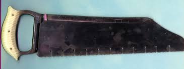
I'm sure you know how a saw sounds. Schrrck-schrrck-schrrck-schrrck. Usually we have one person sawing and the other four trying to hold the cadavre still. Sometimes we have everything dissected away and are right down on the bone, and sometimes we're just flat out sawing through the face or some such thing. That's always a little weird feeling.
A lot of other groups like to use chisels. I know this because you can hear a dozen chisels clinking away throughout the room. If you stop to listen, it sounds just like shop class. Chink, chink, schrrck-schrrck-schrrck, chink, clunk, clink, chink!
That said, what is it we're actually doing? As much as it might sound like it, we're not just tearing the face apart. The first sawing/chiseling we had to do was so we could get a good look at the jaw, and later we carefully took out a section of the muzzle, but the best was definitely when it was time to look at the brain. Despite having giant, unwieldy saws, we needed to plan and cut very carefully. You probably noticed that skulls are round, not square, and it's definitely not a good idea to start sawing into the brain. When we finally prised off the cranium, we learned that we had to then go hack off bone around the spinal cord so we could actually cut the brain stem and get the brain out. It was a long day.
At the end of the day, as normal and reasonable as it may have been at the time, it just does not do to try and tell your non-vet friends how you sawed a dog's face in half today.
Several times now, we've had to cut through bones in anatomy lab to get to what we want to look at. The first time was in the thorax, where we had to break off the ribs so we could look inside and see the heart and lungs and stuff. This involved using a pair of giant wire cutters ("bone cutters," sure) to snap the ribs at both ends with a loud crack. We left the muscles and everything in place, so that the thoracic wall makes a little flap that we can lift to peek into the thorax. How cute.
We only needed to go through bone one other time last semester, to see the spinal cord, and the rest of the bone sawing has been happening this semester. You see, this semester, we've spent seven weeks just looking at the head. There's a lot to see! Unfortunately a lot happens to be inside the head.
The thing that really made me want to tell a story about all this bone stuff is the noise. I already described the wire cutter bone cutters, but clearly that's not going to work very well for something like a skull. Our other options are saws and chisels.
My group likes to use the saw. It's completely barbaric feeling, but it gets the job done. The saws we have are these square things about as long as your forearm, that aren't too far off from this frightening picture I got from google:
I'm sure you know how a saw sounds. Schrrck-schrrck-schrrck-schrrck. Usually we have one person sawing and the other four trying to hold the cadavre still. Sometimes we have everything dissected away and are right down on the bone, and sometimes we're just flat out sawing through the face or some such thing. That's always a little weird feeling.
A lot of other groups like to use chisels. I know this because you can hear a dozen chisels clinking away throughout the room. If you stop to listen, it sounds just like shop class. Chink, chink, schrrck-schrrck-schrrck, chink, clunk, clink, chink!
That said, what is it we're actually doing? As much as it might sound like it, we're not just tearing the face apart. The first sawing/chiseling we had to do was so we could get a good look at the jaw, and later we carefully took out a section of the muzzle, but the best was definitely when it was time to look at the brain. Despite having giant, unwieldy saws, we needed to plan and cut very carefully. You probably noticed that skulls are round, not square, and it's definitely not a good idea to start sawing into the brain. When we finally prised off the cranium, we learned that we had to then go hack off bone around the spinal cord so we could actually cut the brain stem and get the brain out. It was a long day.
At the end of the day, as normal and reasonable as it may have been at the time, it just does not do to try and tell your non-vet friends how you sawed a dog's face in half today.
Subscribe to:
Posts (Atom)