Warning: Posts relating to necropsy week are generally not for the faint of heart.
When we do a post-mortem examination, naturally we have to record what we found, which involves making notes on a blue sheet with boxes for each body system. My classmates tend to do this afterwards, over lunch, which is probably pretty sensible, as you'll see. I'm too paranoid that I'm going to completely forget something important, so I have to write things down as I think of them.
Today, once we removed a section of ribs, we set them down to one side and got to work removing the heart and lungs. As an aside, we had a really fascinating case. The dog had suffered from a bloody nose for about a month, which had gotten worse until the owners decided to euthanise. The abdomen was looking a bit "pot bellied" as well, but the dog was perfectly bright, happy, and eating well. However, when we opened the abdomen, a massive liver tumour practically jumped out at us. When we checked the nasal cavity (by sawing the face in half through the nose, by the way), it turned out to have, as suspected, and totally unrelated, a nasal tumour as well. Unlucky dog, eh?
So while we were busy getting excited about all the pathology, it turns out we had set the slab of ribs and muscles and fat right on top of our blue sheet. Whoops. Eventually, we noticed this, and when we picked the ribs up, the sheet went with it. I tried to pull the sheet off, but it wouldn't come. The fat was stuck fast. Pull pull pull, yank yank yank. Ummm, there's a giant mass of flesh stuck to half our paper.
Somehow, I managed to free our sheet without tearing it, leaving a large wet area around one quadrant. Blood and goop is a pretty normal part of a pathologist's life, so it probably doesn't matter apart from me feeling self-conscious. I proceeded to make the sheet worse and worse through the course of the lab by writing down notes and measurements with increasingly bloody gloves.
My partner and I looked at each other at the end of the morning and debated whether we should rewrite it, but there wasn't much time before the afternoon tutorial, so we figured, "Nahh, it probably doesn't matter." It planted the seed of doubt, however, and after the tutorial had finished, we both started thinking, "Well, maaaaaybeee we should rewrite it. Everyone else's PM reports are all pristine and neat." However, after a brief search, we discovered the clinician had already taken our pile of papers. Oh well.
Showing posts with label Working With Dead Things. Show all posts
Showing posts with label Working With Dead Things. Show all posts
Monday, 29 September 2014

Wednesday, 12 March 2014
Brain Tally
On the whiteboard in the pathology museum:
A: 1/4 1
H: 1
L: 0
A: 0.2
G: 1/2 + 1
L: 1
You see, we're supposed to remove as many brains as possible over the week, and the prof is keeping score. For the record, my 1/4 was from a kitten brain, because the skull was so tiny it was basically snip-snip with the scissors, rather than a big ordeal with a saw.
A: 1/4 1
H: 1
L: 0
A: 0.2
G: 1/2 + 1
L: 1
You see, we're supposed to remove as many brains as possible over the week, and the prof is keeping score. For the record, my 1/4 was from a kitten brain, because the skull was so tiny it was basically snip-snip with the scissors, rather than a big ordeal with a saw.
Tuesday, 11 March 2014
My Life is A Horror Movie
Warning: I'm on necropsy this week. It's a little hard to tell, because we get so desensitised that we can easily eat our lunch while reviewing pictures of bloody, disemboweled animal corpses, but I'm pretty sure that talk of necropsy is disturbing imagery for the average person. Proceed with caution. The point of the post is, basically, blood and gore.
Here's how necropsy rotation works:
We show up in the morning (9am start! how unusual!) and have a little tutorial with the pathologist teaching the roster. We go over all the cases that have been submitted for the day and talk about our differential diagnoses and what we might expect to find on post-mortem. We divvy up the cases between the students, then head off to the PM room floor.
The PM room is designed with an elevated viewing area in front of the entrance, which includes things like the tutorial room and locker room doors, and a lower floor area that takes up most of the room. You have to go down some steps and wade through disinfectant to get there, and the floor is usually pretty wet. It's filled with drains, metal tables, and all the equipment you might want: tubs to carry organs around in, clamps to put heads in while you saw them open, tables of knives, a band saw in one corner, stuff like that.
Something slightly distressing to some is that the animals don't always arrive dead. The upshot is that means a very fresh PM, rather than something that's been in a freezer for a month, but the downside is we have to euthanise them ourselves. In the small animal world, it's a simple injection. But euthanising large animals is, I find, particularly unpleasant and not for the faint of heart--the larger the animal, the worse it is. They're rendered insensible (and probably dead) by a captive bolt into their skull, then to make extra super sure that they're dead, you slash open their throat so they exsanguinate (translation: ocean of blood on the floor).
Once they're not only merely dead, but really most sincerely dead, the students are unleashed. Basically, you go through everything, outside and inside, in a systematic way. While it's all very scientific and medical, to an outside observer, it's skinning, tearing out the heart and lungs, cutting through vessels and spilling blood everywhere, stuff like that. It's quite hack-and-slash feeling. When it's time to look at the abdomen, you've just got organs all over the place. And to check out the brain, you break out the saws and cut open the skull. The things you didn't realise you signed up for, eh?
One of the most disconcerting things is that the muscles keep twitching due to the nerves firing. I always find it startling, even when I know it's coming, that when you cut through a big nerve (like the sciatic), the leg jerks.
Something a little morbidly interesting is that the method of euthanasia dramatically impacts what the PM is like. I had a calf with a suspected central nervous system disease, so we didn't want to use a captive bolt and destroy the brain. I used the injection instead, which means the calf didn't exsanguinate like the others. Out of the three calves that were PM'd at the same time, guess whose table was drowned in blood?
At the end of the day, we have to write up everything we found and submit it for the pathologist to edit and finalise. We have these green sheets to take notes on, and by the end of the morning, both sheet and pen are saturated with essence of dead animal. Yet we just fold them up, take them home, and type it up on our computers like there's nothing unusual. I guess, to be honest, there isn't.
Here's how necropsy rotation works:
We show up in the morning (9am start! how unusual!) and have a little tutorial with the pathologist teaching the roster. We go over all the cases that have been submitted for the day and talk about our differential diagnoses and what we might expect to find on post-mortem. We divvy up the cases between the students, then head off to the PM room floor.
The PM room is designed with an elevated viewing area in front of the entrance, which includes things like the tutorial room and locker room doors, and a lower floor area that takes up most of the room. You have to go down some steps and wade through disinfectant to get there, and the floor is usually pretty wet. It's filled with drains, metal tables, and all the equipment you might want: tubs to carry organs around in, clamps to put heads in while you saw them open, tables of knives, a band saw in one corner, stuff like that.
Something slightly distressing to some is that the animals don't always arrive dead. The upshot is that means a very fresh PM, rather than something that's been in a freezer for a month, but the downside is we have to euthanise them ourselves. In the small animal world, it's a simple injection. But euthanising large animals is, I find, particularly unpleasant and not for the faint of heart--the larger the animal, the worse it is. They're rendered insensible (and probably dead) by a captive bolt into their skull, then to make extra super sure that they're dead, you slash open their throat so they exsanguinate (translation: ocean of blood on the floor).
Once they're not only merely dead, but really most sincerely dead, the students are unleashed. Basically, you go through everything, outside and inside, in a systematic way. While it's all very scientific and medical, to an outside observer, it's skinning, tearing out the heart and lungs, cutting through vessels and spilling blood everywhere, stuff like that. It's quite hack-and-slash feeling. When it's time to look at the abdomen, you've just got organs all over the place. And to check out the brain, you break out the saws and cut open the skull. The things you didn't realise you signed up for, eh?
One of the most disconcerting things is that the muscles keep twitching due to the nerves firing. I always find it startling, even when I know it's coming, that when you cut through a big nerve (like the sciatic), the leg jerks.
Something a little morbidly interesting is that the method of euthanasia dramatically impacts what the PM is like. I had a calf with a suspected central nervous system disease, so we didn't want to use a captive bolt and destroy the brain. I used the injection instead, which means the calf didn't exsanguinate like the others. Out of the three calves that were PM'd at the same time, guess whose table was drowned in blood?
At the end of the day, we have to write up everything we found and submit it for the pathologist to edit and finalise. We have these green sheets to take notes on, and by the end of the morning, both sheet and pen are saturated with essence of dead animal. Yet we just fold them up, take them home, and type it up on our computers like there's nothing unusual. I guess, to be honest, there isn't.
Monday, 10 March 2014
Necropsy, From the Tiny to the Giant
Warning: Descriptions of dead animals.
Pathology rounds are every friday afternoon, and for as long as we've been in vet school, we've been encouraged to attend. It requires nothing on your part, simply show up, stand around the post-mortem room, and watch. At first, it's practically incomprehensible, but as you progress through the years you start understanding more and more of what's being said and what you're looking at. Pathology rounds are a regular fixture in (some of) our lives, a weekly occurrence that's gone on as long as vet school has.
And finally, it becomes our turn to take the floor, our turn to stand on the other side of the room and hold up the cold entrails of dead animals for all to see.
You see, this week I'm on necropsy. On friday, we are the ones to present the cases that come in during the week. Some come in dead already, some arrive alive and get euthanised on the premises. We hack apart their bodies completely and utterly, send samples off to the lab, and save whatever is interesting for path rounds.
Today we had three animals: a kitten, an alpaca, and a cow. I got the kitten. Nine days old, it had been found dead underneath its mom, suspected crushed/smothered. Everything was so tiny, it was hard to do the PM steps like normal. Her teeny tiny mouth and tongue, and teeny tiny GIT and kidneys and spleen and everything, and super teeny tiny uterus--actually it was pretty cute. Or at least I thought so, and one other person, but the production trackers thought that was weird, and were all, "Psh, smallies people." But at any rate, I kept accidentally crushing and tearing things, and fur got all up in everything, and it was very difficult to cut organs open and get a good look at everything. I practically needed a microscope.
So here I am, with my animal that can fit in the palm of my hand, holding up its tiny heart and lungs between two fingers and poking at it with a pair of forceps, and at the table next to me is the team working on the cow: heart the size of my head, a mountain of rumen contents all over the floor, sawing and hacking with giant knives, cutting the ribs with massive bolt cutters, an ocean of blood leaking out of their animal. The liver alone was an entire armful. It amazes me how these two very different creatures are so similar in their composition. My kitten's little heart the size of a fingernail had all the same components as the massive cow heart, just miniature.
The alpaca was interesting, too, just by way of being an alpaca.
Pathology rounds are every friday afternoon, and for as long as we've been in vet school, we've been encouraged to attend. It requires nothing on your part, simply show up, stand around the post-mortem room, and watch. At first, it's practically incomprehensible, but as you progress through the years you start understanding more and more of what's being said and what you're looking at. Pathology rounds are a regular fixture in (some of) our lives, a weekly occurrence that's gone on as long as vet school has.
And finally, it becomes our turn to take the floor, our turn to stand on the other side of the room and hold up the cold entrails of dead animals for all to see.
You see, this week I'm on necropsy. On friday, we are the ones to present the cases that come in during the week. Some come in dead already, some arrive alive and get euthanised on the premises. We hack apart their bodies completely and utterly, send samples off to the lab, and save whatever is interesting for path rounds.
Today we had three animals: a kitten, an alpaca, and a cow. I got the kitten. Nine days old, it had been found dead underneath its mom, suspected crushed/smothered. Everything was so tiny, it was hard to do the PM steps like normal. Her teeny tiny mouth and tongue, and teeny tiny GIT and kidneys and spleen and everything, and super teeny tiny uterus--actually it was pretty cute. Or at least I thought so, and one other person, but the production trackers thought that was weird, and were all, "Psh, smallies people." But at any rate, I kept accidentally crushing and tearing things, and fur got all up in everything, and it was very difficult to cut organs open and get a good look at everything. I practically needed a microscope.
So here I am, with my animal that can fit in the palm of my hand, holding up its tiny heart and lungs between two fingers and poking at it with a pair of forceps, and at the table next to me is the team working on the cow: heart the size of my head, a mountain of rumen contents all over the floor, sawing and hacking with giant knives, cutting the ribs with massive bolt cutters, an ocean of blood leaking out of their animal. The liver alone was an entire armful. It amazes me how these two very different creatures are so similar in their composition. My kitten's little heart the size of a fingernail had all the same components as the massive cow heart, just miniature.
The alpaca was interesting, too, just by way of being an alpaca.
Friday, 20 December 2013
Week of Awesome Does Not Disappoint, Part 2
Warning: Standard warning about a post that describes more graphic procedures and/or dead animals. This one has some pictures. Though they are the tamer ones, read with discretion.
Dentistry: Day 2 was dentistry. That's right, everything in part 1 was just tuesday. Dentistry is actually less horrible than you'd expect, although the high-pitched whine of the ultrasonic scaler is still slightly distressing.
Basically, we took out teeth until we got tired of it. Depending on how many roots a tooth has, it's more or less complex, but they all involve sticking a "dental elevator" down into the tooth socket and pushing and rotating until the tooth jiggles out. Only one of the drills was working, so there was a rather awkward queue because everyone lifted up their table with the dead dog on it, and carried it over (because the drill is on a short power cord). So at any given point, there was a group using the drill, and a group next to them that had brought over their giant metal table.
Dogs don't really get cavities. We were actually using the drill for extractions. You use it to slice the tooth apart between the roots, and also to expose the roots from under the bone. The other thing we did was to practice local blocks. As most of the nerves come out of specific holes in the skull, the aim was to feel for those holes and see if we could stick the needle into them. The result was a dog mouth full of needles.
Orthopaedics: Half of day 3 was dedicated to the wonderful world of metal bone implants. Drills, pins, plates, screws, wire, interlocking nails, prosthetic nylon ligaments. There were too many things to practice so each group only picked a few things. Depending on what you picked, the surgeon would come over with the bone saw and make a fracture in your bone of choice.
The first thing my partner and I did was an ulnar fracture. This involved two pins down the shaft of the bone for stability, and a wire inserted in a figure eight to create tension.


Ophthalmology: The last thing we did was eye and eyelid surgery, with a boarded ophthalmologist. It's kind of weird and creepy, but it's a lot more comforting to do it on a dead dog, where you know it doesn't matter even if you somehow exploded the eyeball or lost it or something. The downside is that since we only had one eye each to work with, a whole bunch of lid surgeries on one lid tend to make it look like franken-dog. The most exciting part was cutting or sewing onto the cornea, because it's amazing that you can even do that. It's layered like an onion, so if you have to remove a diseased piece, you cut a little square and it just peels off. Another cool thing you can do is actually use the conjunctiva like a bandage, where you cut a little pedicle flap out of the surrounding membranes, bring it across the eye, and sew it onto the cornea. I wanted to include the picture of this, but dead dog eye surgery is really creepy so I thought better of it.
Also, the lens is surprisingly soft and pliant. You think of it as a hard thing, but it isn't.

Dentistry: Day 2 was dentistry. That's right, everything in part 1 was just tuesday. Dentistry is actually less horrible than you'd expect, although the high-pitched whine of the ultrasonic scaler is still slightly distressing.
Basically, we took out teeth until we got tired of it. Depending on how many roots a tooth has, it's more or less complex, but they all involve sticking a "dental elevator" down into the tooth socket and pushing and rotating until the tooth jiggles out. Only one of the drills was working, so there was a rather awkward queue because everyone lifted up their table with the dead dog on it, and carried it over (because the drill is on a short power cord). So at any given point, there was a group using the drill, and a group next to them that had brought over their giant metal table.
Dogs don't really get cavities. We were actually using the drill for extractions. You use it to slice the tooth apart between the roots, and also to expose the roots from under the bone. The other thing we did was to practice local blocks. As most of the nerves come out of specific holes in the skull, the aim was to feel for those holes and see if we could stick the needle into them. The result was a dog mouth full of needles.
Orthopaedics: Half of day 3 was dedicated to the wonderful world of metal bone implants. Drills, pins, plates, screws, wire, interlocking nails, prosthetic nylon ligaments. There were too many things to practice so each group only picked a few things. Depending on what you picked, the surgeon would come over with the bone saw and make a fracture in your bone of choice.
The first thing my partner and I did was an ulnar fracture. This involved two pins down the shaft of the bone for stability, and a wire inserted in a figure eight to create tension.
Then I did a knee repair of a ruptured cranial cruciate ligament (in people referred to as an ACL). Since it wasn't actually ruptured, I had to go into the joint and cut the ligament, but that's normally part of the procedure since you have to inspect the joint anyway. Then you drill holes into the tibia that are used to hold the nylon prosthesis into a position that approximates the function of the ligament. The nylon loops around a bit of bone in the back, goes around the tibia via the holes, and voila.
My classmates got up to some other exciting stuff, like external skeletal fixation, where the implants come out of the skin and get connected by bars. They look pretty impressive.
Ophthalmology: The last thing we did was eye and eyelid surgery, with a boarded ophthalmologist. It's kind of weird and creepy, but it's a lot more comforting to do it on a dead dog, where you know it doesn't matter even if you somehow exploded the eyeball or lost it or something. The downside is that since we only had one eye each to work with, a whole bunch of lid surgeries on one lid tend to make it look like franken-dog. The most exciting part was cutting or sewing onto the cornea, because it's amazing that you can even do that. It's layered like an onion, so if you have to remove a diseased piece, you cut a little square and it just peels off. Another cool thing you can do is actually use the conjunctiva like a bandage, where you cut a little pedicle flap out of the surrounding membranes, bring it across the eye, and sew it onto the cornea. I wanted to include the picture of this, but dead dog eye surgery is really creepy so I thought better of it.
Also, the lens is surprisingly soft and pliant. You think of it as a hard thing, but it isn't.
Unfortunately, as I was squishing it gently in amazement, I broke it and it burst apart into a pile of clear, gelatinous goo. Whoops. The eruption of laughter at another table a few minutes later indicated that other students encountered this unexpected turn of events as well.
Wednesday, 18 December 2013
Week of Awesome Does Not Disappoint, Part 1
Warning: This post discusses medical procedures on dead dogs. Neither aspect of that sentence is very pretty, so if you're squeamish, or sensitive about animals, proceed with caution.
As the title suggests, this week has been heaps of fun. Starting tuesday morning, we got our cadavres, and had three packed-full days to get as much out of them as possible. We cut, stabbed, tied, dissected, and tubed every single part of their little bodies.
The dogs come from a nearby shelter, usually a variable amount less than ten, and the roster is kept small so that there can be two students to a dog. In first year, it was five students to a dog, and in fourth year it was groups of three for surgery lab, so this is a huge opportunity. Somewhat distressingly, the shelter purportedly never has an issue with supplying enough dogs, and the animals are not withheld at the shelter for more than a day. Most of the animals are pit-bull crosses, so I think there's an unfortunate excess of unwanted, potentially dangerous young animals in the area. Anyway, we had 8 dogs for 15 students, and when they first arrived we had to be speedy to take advantage of certain things before rigor mortis set in.
First thing we had to do was put a little bottle in their mouths to keep their jaws open. Believe it or not, it's very difficult to later do stomach tubing if their jaw is locked closed. The next step was to jump to the procedures that involved fluids: blood, joint fluid, CSF, and bone marrow. For every procedure, the vets gave a demonstration, and we went off and performed it on our own dogs. Here are some of the highlights.
CSF tap: To obtain cerebrospimal fluid, you stick a needle in between the vertebrae into the space around the spinal cord. You can do this right behind the skull, or in the lower back. After you feel your bony landmarks, you basically just go for it, and pop the needle in. Unless you are awesome(*), it likely will hit one of the bones on either side and needs to be redirected--and let me tell you, the feel of a needle crunching against bone is horrible. When you get it into the joint space, it goes all the way in, nice and smooth. When you don't, it smacks into bone early and won't move, so you have to poke, redirect, poke, and redirect until you get it, with an awful scraping sensation against the not-quite-solid surface.
*I got the lumbosacral cistern on the first try. That one has a slower flow in general, and also our dogs are dead, so I wasn't sure at first. But I didn't want to give up on it, so I waited, and 30 seconds later came the satisfying clear goop oozing out of the hub of my needle.
Bone marrow aspirate: Take what I was saying about bone before, now imagine a giant metal stick with a handle that you shove forcibly down a shoulder into the humerus. Surprisingly good for getting out stress, as you crank that handle back and forth like you're grinding your fist into your worst enemy's face. If you get a little too into it, your marrow aspirate turns into a core biopsy, because you punch through the cortex and get a full piece of bone, rather than sucking up the viscous marrow. There is actually a difference in what they tell you diagnostically.
Chest tube: This one is both terrifying and hilarious, as your big, thick chest tube goes onto a similarly big, thick metal stylette with a pointy end. You grab the bottom about a centimeter up from the pointy end, to avoid skewering the patient, and with your other arm you bang it through the chest wall with one swift smack. Pop! Pointy end into the patient, and your fist stops you from stabbing the heart or something.
Another fun aspect is that you need to make a tunnel under the skin so you don't accidentally let a bunch of air into the chest. This means an assistant grabs fistfulls of dog fur and holds them to the side, so that once the chest tube is in, when they let go the loose skin will fall back into place and make it so the entry point in the skin was several rib spaces farther down than the point through the body wall. When you're all done, you can really see the tube there, right under the skin, a smooth squishy cylinder. You secure the tube in place by what is known as the Chinese finger trap suture, not even kidding.
Oesophagostomy: Since we had the foresight to prop open our dog's mouth, we were able to attempt stomach tubes. There are a few methods, like through the nose or through the oesophagus. The latter is useful because it can be a semi-permanent way to bypass the mouth or provide/force nutrition easily. The way it works is you make a cut on their neck, into the oesophagus, and stick the tube in from the outside. You have to fiddle around, grabbing the tube from the inside and fussing with it until it's in the direction you need, and apparently this is next to impossible in a dead dog. But because, as we've already established, I am awesome, I totally got ours into place.
Exploratory laparotomy and experimental surgery: Towards the end of the first day, we had to gut the dogs so they wouldn't become too smelly and horrid over the week. But there's no sense in just gutting them, so we explored their abdomens first. We checked out all the organs, took biopsies of everything (which involve a number of bizarre stabby instruments), did pretend surgery on the intestines, and generally poked, prodded, and sliced anything we could get our hands on. The pretend intestine surgery is fun, because you cut out a piece, tie it back together, then turn it into a water balloon to see if your seal was water-tight--that is, hold it off and inject it with water. We even castrated our dead dog, and put in a urinary catheter such that we could see when it got to the bladder.
"If you get bored": As if we didn't have enough to do, they had tons of extra equipment to make extra sure we didn't run into any down time. There were otoscopes, slides for looking at our samples, nail clippers, syringe and needles for lymph node aspirates, you name it. Unfortunately, because our dogs arrived an hour later than they were supposed to, we ended up quite tight for time.
As the title suggests, this week has been heaps of fun. Starting tuesday morning, we got our cadavres, and had three packed-full days to get as much out of them as possible. We cut, stabbed, tied, dissected, and tubed every single part of their little bodies.
The dogs come from a nearby shelter, usually a variable amount less than ten, and the roster is kept small so that there can be two students to a dog. In first year, it was five students to a dog, and in fourth year it was groups of three for surgery lab, so this is a huge opportunity. Somewhat distressingly, the shelter purportedly never has an issue with supplying enough dogs, and the animals are not withheld at the shelter for more than a day. Most of the animals are pit-bull crosses, so I think there's an unfortunate excess of unwanted, potentially dangerous young animals in the area. Anyway, we had 8 dogs for 15 students, and when they first arrived we had to be speedy to take advantage of certain things before rigor mortis set in.
First thing we had to do was put a little bottle in their mouths to keep their jaws open. Believe it or not, it's very difficult to later do stomach tubing if their jaw is locked closed. The next step was to jump to the procedures that involved fluids: blood, joint fluid, CSF, and bone marrow. For every procedure, the vets gave a demonstration, and we went off and performed it on our own dogs. Here are some of the highlights.
CSF tap: To obtain cerebrospimal fluid, you stick a needle in between the vertebrae into the space around the spinal cord. You can do this right behind the skull, or in the lower back. After you feel your bony landmarks, you basically just go for it, and pop the needle in. Unless you are awesome(*), it likely will hit one of the bones on either side and needs to be redirected--and let me tell you, the feel of a needle crunching against bone is horrible. When you get it into the joint space, it goes all the way in, nice and smooth. When you don't, it smacks into bone early and won't move, so you have to poke, redirect, poke, and redirect until you get it, with an awful scraping sensation against the not-quite-solid surface.
*I got the lumbosacral cistern on the first try. That one has a slower flow in general, and also our dogs are dead, so I wasn't sure at first. But I didn't want to give up on it, so I waited, and 30 seconds later came the satisfying clear goop oozing out of the hub of my needle.
Bone marrow aspirate: Take what I was saying about bone before, now imagine a giant metal stick with a handle that you shove forcibly down a shoulder into the humerus. Surprisingly good for getting out stress, as you crank that handle back and forth like you're grinding your fist into your worst enemy's face. If you get a little too into it, your marrow aspirate turns into a core biopsy, because you punch through the cortex and get a full piece of bone, rather than sucking up the viscous marrow. There is actually a difference in what they tell you diagnostically.
Chest tube: This one is both terrifying and hilarious, as your big, thick chest tube goes onto a similarly big, thick metal stylette with a pointy end. You grab the bottom about a centimeter up from the pointy end, to avoid skewering the patient, and with your other arm you bang it through the chest wall with one swift smack. Pop! Pointy end into the patient, and your fist stops you from stabbing the heart or something.
Another fun aspect is that you need to make a tunnel under the skin so you don't accidentally let a bunch of air into the chest. This means an assistant grabs fistfulls of dog fur and holds them to the side, so that once the chest tube is in, when they let go the loose skin will fall back into place and make it so the entry point in the skin was several rib spaces farther down than the point through the body wall. When you're all done, you can really see the tube there, right under the skin, a smooth squishy cylinder. You secure the tube in place by what is known as the Chinese finger trap suture, not even kidding.
Oesophagostomy: Since we had the foresight to prop open our dog's mouth, we were able to attempt stomach tubes. There are a few methods, like through the nose or through the oesophagus. The latter is useful because it can be a semi-permanent way to bypass the mouth or provide/force nutrition easily. The way it works is you make a cut on their neck, into the oesophagus, and stick the tube in from the outside. You have to fiddle around, grabbing the tube from the inside and fussing with it until it's in the direction you need, and apparently this is next to impossible in a dead dog. But because, as we've already established, I am awesome, I totally got ours into place.
Exploratory laparotomy and experimental surgery: Towards the end of the first day, we had to gut the dogs so they wouldn't become too smelly and horrid over the week. But there's no sense in just gutting them, so we explored their abdomens first. We checked out all the organs, took biopsies of everything (which involve a number of bizarre stabby instruments), did pretend surgery on the intestines, and generally poked, prodded, and sliced anything we could get our hands on. The pretend intestine surgery is fun, because you cut out a piece, tie it back together, then turn it into a water balloon to see if your seal was water-tight--that is, hold it off and inject it with water. We even castrated our dead dog, and put in a urinary catheter such that we could see when it got to the bladder.
"If you get bored": As if we didn't have enough to do, they had tons of extra equipment to make extra sure we didn't run into any down time. There were otoscopes, slides for looking at our samples, nail clippers, syringe and needles for lymph node aspirates, you name it. Unfortunately, because our dogs arrived an hour later than they were supposed to, we ended up quite tight for time.
Thursday, 11 July 2013
What did you do today?
Standard warning: potentially unpleasant graphic concepts
Thursdays are lab days. Sometimes they're long, or a bit dull, or kind of cool. And then sometimes they're like today.
In the morning is the small animal lab, either medicine or surgery. Today we did dentistry. If you've ever had your pet get a dental from your vet, that's what we practised, and it's actually not very different from when you go to the dentist yourself. We use all the same tools, including the ultrasonic scaler which is the thing that makes the noise and uses the water spray while it scrapes against the teeth. So that was pretty neat, checking out all the teeth and cleaning off the tartar--the noise isn't quite so bad when you're on the other end of it.
The part about this lab that made it a rather unique experience is something you may have already thought of: what, exactly, were we practising on? Dogs. Dog heads, of course. Severed dog heads. We show up to lab and there is a huge pile of dog heads, and I go over and pick one out for me and my partner and carry it back to our table. So we just have this dog head (a pretty cute dog to be honest), that we are lifting up and rolling around as needed, prying open the jaws and cleaning its dead teeth. I have to say, that is a bit bizarre--cleaning a dead dog's teeth.
The other thing we did was practise extractions, which is a bit more of an ordeal than in people, because dog teeth have massive roots. It's more of a surgery, where you have to slice into the gums and make a flap, so you can get your tools way down to the end of the root. This involved some fun instruments like the drill (just like in people, if you've ever had a cavity done), and a lot of prying and leveraging to try and shove the tooth out. Like my human dentist once told me, it's not about strength, it's all about technique--you have to push in the right way, and the right direction. And it's actually pushing, not pulling.
That was in the morning. In the afternoon we have a large animal lab, either cows or sheep. Today it was sheep.
We've been learning about investigating ill-thrift in sheep, and there is a farm that is working with the university to investigate a problem in their flock. They have a whole bunch of very skinny sad looking ewes, and we the fourth years are going to be the ones helping to find out what's wrong.
Our labs are divided into half the class, so we have 50 people, and we were working in pairs. That means 25 sheep or so waiting for us in lab, and the first thing we did was the Great Sheep Massacre. We queued up, each pair with their sheep, and had a rather rapid-fire, bloody, sheep killing spree. We used captive bolts, which fires a bolt into their brain to destroy their pain perception and render them unconscious instantly, and then cut their throats to make certain they were dead (though theoretically the captive bolt kills them).
I partnered up with a very very quiet girl in my class. I did the captive bolt, which is a deceptively heavy item to wield. That involved putting the thing on the right spot on the sheep's head, then pressing the button. BANG, sheep collapses. My partner cut the throat, which involves twisting the sheep's neck around your boot and hacking through all the muscles and trachea until you get a massive fountain/lake of blood, and I have to say for such a quiet meek seeming girl, my partner had absolutely no trouble with this. Once the deed was done (which only took a few seconds), we quickly got out of the way and handed off the tools to the next group. We dragged off our sheep to an open spot in the floor, adding to the dozen bloody trails along the floor. I can't imagine what it must have looked like if some random person from the public had shown up. Dead sheep all across the floor, wide bloody trails behind them, a sea of blood at the centres of two ritualistic sheep-killing death spots, students queued up with sheep at each.
Once that was all over we proceeded to the post-mortems. As these are all very sickly, emaciated sheep, we went through our protocol of checking out each organ system in the hopes of finding a consistent culprit. Common boring ones include teeth and feet. I call these boring because you don't even have to open your dead sheep in order to get a diagnosis. Apart from those things, we had to check out liver, lungs, and intestines, looking for typical lesions of the diseases we've studied. The aim is that every pair writes up their final diagnosis on a board, and we'll get sent all the results. We'll analyse them, see if there's a common cause, and then write up a report about it.
Our particular sheep turned out to not be very exciting. I thought we had some lung lesions, but it turned out they weren't significant enough to call it pneumonia. We ended up not finding any lesions, which suggests either malnutrition, or maybe parasites. Which brings me to the next part.
We did something called a "worm count." When animals have parasites in their intestines, you can count the eggs released in their poop, but it's much more accurate to count the worms themselves. You take out parts of the intestinal tract, and then go through a fancy procedure to wash out all the contents into a bucket, dilute the worms, and put them onto a white tray to count them. It's winter here, and cold outside, and we had cold water, and I did not have a fun time. I spent forever running meters upon meters of intestine through my fingers in a bucket of cold water, to help get the worms off and into the water. By the end of the lab I could hardly feel my hands. I was trying to pick up microscopic worms and put them onto a slide, and my goodness my fingers could barely move. In the end, our sheep didn't have any parasites. After all that.
So our sheep ended up with a diagnosis of "unknown," and we attributed the emaciation to malnourishment just as the default. Once we get everybody's results, we'll see if any particular disease emerged as statistically significant. A few people did have some knobbly livers and diseased intestines, so it will be interesting to see the final results. I believe this is an actual investigation, and the professors are going to relay the information to the farmer, so it's pretty cool to be a part of that.
Thursdays are lab days. Sometimes they're long, or a bit dull, or kind of cool. And then sometimes they're like today.
In the morning is the small animal lab, either medicine or surgery. Today we did dentistry. If you've ever had your pet get a dental from your vet, that's what we practised, and it's actually not very different from when you go to the dentist yourself. We use all the same tools, including the ultrasonic scaler which is the thing that makes the noise and uses the water spray while it scrapes against the teeth. So that was pretty neat, checking out all the teeth and cleaning off the tartar--the noise isn't quite so bad when you're on the other end of it.
The part about this lab that made it a rather unique experience is something you may have already thought of: what, exactly, were we practising on? Dogs. Dog heads, of course. Severed dog heads. We show up to lab and there is a huge pile of dog heads, and I go over and pick one out for me and my partner and carry it back to our table. So we just have this dog head (a pretty cute dog to be honest), that we are lifting up and rolling around as needed, prying open the jaws and cleaning its dead teeth. I have to say, that is a bit bizarre--cleaning a dead dog's teeth.
The other thing we did was practise extractions, which is a bit more of an ordeal than in people, because dog teeth have massive roots. It's more of a surgery, where you have to slice into the gums and make a flap, so you can get your tools way down to the end of the root. This involved some fun instruments like the drill (just like in people, if you've ever had a cavity done), and a lot of prying and leveraging to try and shove the tooth out. Like my human dentist once told me, it's not about strength, it's all about technique--you have to push in the right way, and the right direction. And it's actually pushing, not pulling.
That was in the morning. In the afternoon we have a large animal lab, either cows or sheep. Today it was sheep.
We've been learning about investigating ill-thrift in sheep, and there is a farm that is working with the university to investigate a problem in their flock. They have a whole bunch of very skinny sad looking ewes, and we the fourth years are going to be the ones helping to find out what's wrong.
Our labs are divided into half the class, so we have 50 people, and we were working in pairs. That means 25 sheep or so waiting for us in lab, and the first thing we did was the Great Sheep Massacre. We queued up, each pair with their sheep, and had a rather rapid-fire, bloody, sheep killing spree. We used captive bolts, which fires a bolt into their brain to destroy their pain perception and render them unconscious instantly, and then cut their throats to make certain they were dead (though theoretically the captive bolt kills them).
I partnered up with a very very quiet girl in my class. I did the captive bolt, which is a deceptively heavy item to wield. That involved putting the thing on the right spot on the sheep's head, then pressing the button. BANG, sheep collapses. My partner cut the throat, which involves twisting the sheep's neck around your boot and hacking through all the muscles and trachea until you get a massive fountain/lake of blood, and I have to say for such a quiet meek seeming girl, my partner had absolutely no trouble with this. Once the deed was done (which only took a few seconds), we quickly got out of the way and handed off the tools to the next group. We dragged off our sheep to an open spot in the floor, adding to the dozen bloody trails along the floor. I can't imagine what it must have looked like if some random person from the public had shown up. Dead sheep all across the floor, wide bloody trails behind them, a sea of blood at the centres of two ritualistic sheep-killing death spots, students queued up with sheep at each.
Once that was all over we proceeded to the post-mortems. As these are all very sickly, emaciated sheep, we went through our protocol of checking out each organ system in the hopes of finding a consistent culprit. Common boring ones include teeth and feet. I call these boring because you don't even have to open your dead sheep in order to get a diagnosis. Apart from those things, we had to check out liver, lungs, and intestines, looking for typical lesions of the diseases we've studied. The aim is that every pair writes up their final diagnosis on a board, and we'll get sent all the results. We'll analyse them, see if there's a common cause, and then write up a report about it.
Our particular sheep turned out to not be very exciting. I thought we had some lung lesions, but it turned out they weren't significant enough to call it pneumonia. We ended up not finding any lesions, which suggests either malnutrition, or maybe parasites. Which brings me to the next part.
We did something called a "worm count." When animals have parasites in their intestines, you can count the eggs released in their poop, but it's much more accurate to count the worms themselves. You take out parts of the intestinal tract, and then go through a fancy procedure to wash out all the contents into a bucket, dilute the worms, and put them onto a white tray to count them. It's winter here, and cold outside, and we had cold water, and I did not have a fun time. I spent forever running meters upon meters of intestine through my fingers in a bucket of cold water, to help get the worms off and into the water. By the end of the lab I could hardly feel my hands. I was trying to pick up microscopic worms and put them onto a slide, and my goodness my fingers could barely move. In the end, our sheep didn't have any parasites. After all that.
So our sheep ended up with a diagnosis of "unknown," and we attributed the emaciation to malnourishment just as the default. Once we get everybody's results, we'll see if any particular disease emerged as statistically significant. A few people did have some knobbly livers and diseased intestines, so it will be interesting to see the final results. I believe this is an actual investigation, and the professors are going to relay the information to the farmer, so it's pretty cool to be a part of that.
Sunday, 30 September 2012
There's Something Fishy About This Lab
A week or two ago, we had a fish handling lab.
Did you know that people anaesthetise fish? Fish are sedated before they are killed commercially, but people also have valuable pet fish or display fish (like in an aquarium) that sometimes get sick and need veterinary care. There are a number of basic diagnostic techniques for investigating what could be wrong with a sick fish, and medicine or even surgery can be used to treat them.
Fish anaesthesia is surprisingly like mammalian anaesthesia. For a dog, the anaesthetic is vaporised and they breathe it in through their lungs via the anaesthetic machine. For a fish, it's basically the same: the drug is dissolved in the water rather than the air, and they "breathe" it in through their gills. If a dog is too deep, you give it pure oxygen to breathe for a while; for a fish, you put them into fresh water to breathe for a while.
Unfortunately for me, I managed to pick crappy fish for my partner and myself. They would not go to sleep. As they get more sedated, they lose their balance reflexes (float sideways and stuff), but it can freak them out a little as they go down. So one would have a panic attack, and that would wake up the other one, which was just drifting off. Then the first one would calm down, but the second one got a fright. The professor came by to increase the dose three times after the first lot! We spent at least half of our lab time staring into the bucket waiting for them to be deep enough to work with. It didn't help that we kept thinking they were fine, picking them up, and then they would flop around and wake up and we'd have to put them back. As an aside, the anaesthetic we were using is derived from clove oil, so the lab smelled like a strange combination of live fish and cloves.
Once they were asleep, we could take them out and do things to them. They survive in the air because their metabolism and respiration is slowed down so much under anaesthesia. Normally you have to keep them really wet to protect their skin, however. Our lab was designed to be a non-recovery lab, using fish at the end of their lives and such, so we didn't worry about that.
First thing we did was take blood. There's a vein in their tail, right in front of the fin, and we spent almost the entire rest of our lab time trying to stab it. If the needle goes through a scale on the way in, you plug up the thing with keratin and have to get a new needle. Little fish have little veins and not a lot of blood, so it was a pretty frustrating endeavour. The other site I tried was the dorsal aorta, which sounds really scary: you stick the needle into the back of their mouths, and that's apparently right where the aorta comes off the heart. I blindly stuck mine in and actually got an entire drop of blood right off the bat, but everyone else I talked to had a much harder time with that one.
Another thing we did was skin scrapes, which is apparently quite useful as fish can get protozoal infections, and if you put the mucus on a slide, you can see the protozoa move around. Our fish were not so interesting.
The last thing we did with the live fish was a gill biopsy. If you don't know anything about fish physiology, I have to tell you, gills are cool. They look cool, they are designed cool, they are just cool. You can also snip off a tiny piece with scissors, put it on a slide, and have a look for disease or degeneration that might be making the fish sick (it would be like people having a lung problem). Electron micrograph photo:

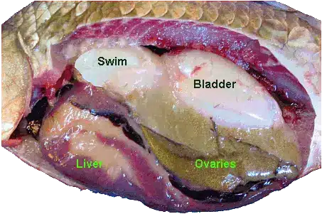
The last thing we did was euthanise the fish and do a post mortem. My partner and I had basically no time to do this, so we only had a quick look before lab ended. They have inflated swim bladders next to the kidney, that was the neatest thing, like a little balloon inside their body cavity. They have teeny tiny little spleens, massive massive ovaries filled with eggs, a simple two chambered heart, and skinny twisty intestines. From google:
Did you know that people anaesthetise fish? Fish are sedated before they are killed commercially, but people also have valuable pet fish or display fish (like in an aquarium) that sometimes get sick and need veterinary care. There are a number of basic diagnostic techniques for investigating what could be wrong with a sick fish, and medicine or even surgery can be used to treat them.
Fish anaesthesia is surprisingly like mammalian anaesthesia. For a dog, the anaesthetic is vaporised and they breathe it in through their lungs via the anaesthetic machine. For a fish, it's basically the same: the drug is dissolved in the water rather than the air, and they "breathe" it in through their gills. If a dog is too deep, you give it pure oxygen to breathe for a while; for a fish, you put them into fresh water to breathe for a while.
Unfortunately for me, I managed to pick crappy fish for my partner and myself. They would not go to sleep. As they get more sedated, they lose their balance reflexes (float sideways and stuff), but it can freak them out a little as they go down. So one would have a panic attack, and that would wake up the other one, which was just drifting off. Then the first one would calm down, but the second one got a fright. The professor came by to increase the dose three times after the first lot! We spent at least half of our lab time staring into the bucket waiting for them to be deep enough to work with. It didn't help that we kept thinking they were fine, picking them up, and then they would flop around and wake up and we'd have to put them back. As an aside, the anaesthetic we were using is derived from clove oil, so the lab smelled like a strange combination of live fish and cloves.
Once they were asleep, we could take them out and do things to them. They survive in the air because their metabolism and respiration is slowed down so much under anaesthesia. Normally you have to keep them really wet to protect their skin, however. Our lab was designed to be a non-recovery lab, using fish at the end of their lives and such, so we didn't worry about that.
First thing we did was take blood. There's a vein in their tail, right in front of the fin, and we spent almost the entire rest of our lab time trying to stab it. If the needle goes through a scale on the way in, you plug up the thing with keratin and have to get a new needle. Little fish have little veins and not a lot of blood, so it was a pretty frustrating endeavour. The other site I tried was the dorsal aorta, which sounds really scary: you stick the needle into the back of their mouths, and that's apparently right where the aorta comes off the heart. I blindly stuck mine in and actually got an entire drop of blood right off the bat, but everyone else I talked to had a much harder time with that one.
Another thing we did was skin scrapes, which is apparently quite useful as fish can get protozoal infections, and if you put the mucus on a slide, you can see the protozoa move around. Our fish were not so interesting.
The last thing we did with the live fish was a gill biopsy. If you don't know anything about fish physiology, I have to tell you, gills are cool. They look cool, they are designed cool, they are just cool. You can also snip off a tiny piece with scissors, put it on a slide, and have a look for disease or degeneration that might be making the fish sick (it would be like people having a lung problem). Electron micrograph photo:
The last thing we did was euthanise the fish and do a post mortem. My partner and I had basically no time to do this, so we only had a quick look before lab ended. They have inflated swim bladders next to the kidney, that was the neatest thing, like a little balloon inside their body cavity. They have teeny tiny little spleens, massive massive ovaries filled with eggs, a simple two chambered heart, and skinny twisty intestines. From google:
Tuesday, 18 September 2012
Being On the Other Side (Part 2)
My job teaching the second year anatomy labs included taking part in the oral examination at the end on topography, which is using external landmarks to identify the location of structures that you can't see. That basically means, if you point anywhere on a dog, I should be able to tell you exactly what is there and what it does. I made a post about it when I had to go through this--it's the first oral exam of the degree!
They use the third year students as scribes. So the professor asks the questions, and the scribe copies down what the student says, so there's some sort of record. This concept was terrifying as an examinee: some older and more knowledgable student hiding in the corner of the room, watching you silently and making notes.
If I had known what it was like behind the scenes, I doubt I would have been so nervous.
For one thing, there is NO space to write. Every student gets one line on the sheet, and each question gets a box (four or five questions each). So I had to condense 2-3 minutes of nervous speed-talking into a box the size of my thumbnail. Well, I guess the details aren't so important after all.
The examiner I was paired with was the other ridiculous factor. He's well known as a ridiculous professor (not in a good way), rather lackadaisical. He was completely relaxed and probably a bit bored, since he had to examine student after student all day long, which was an interesting contrast to the complete terror of the poor second years coming into the room. By the end of the afternoon, he was yawning and joking about beer, as some poor sould who'd been waiting tensely for the entire morning stares at him, pale and waiting for it to all be over. Their minds blanked, they stuttered and misspoke, some went really fast, some had massive pauses. But frankly, most of them were 4/5 or above on every question. The lowest person still scored an overall 70% or so.
What really amazed me was that Prof Lackadaisical wasn't really paying attention. Most of them, the grading process pretty much consisted of the student leaving the room, and he turns to me and says "Yeah, she was fine, 5/5 for everything." As the afternoon stretched on, however, I had to point out things like "Well she DID call this thing this totally-other-thing" and he'd be all "Oh. 4/5 then." Eventually we got to him turning to me and asking "How was that one? Was she right?" and I became the sole arbiter of their grades. So, really, they had nothing to worry about, because it depended on both me and him remembering what the person said, and all the details falling through the cracks in the floor because I had only written like 10% of what they said in the first place. Well, that, and the fact that they all had studied and knew what they were talking about.
They use the third year students as scribes. So the professor asks the questions, and the scribe copies down what the student says, so there's some sort of record. This concept was terrifying as an examinee: some older and more knowledgable student hiding in the corner of the room, watching you silently and making notes.
If I had known what it was like behind the scenes, I doubt I would have been so nervous.
For one thing, there is NO space to write. Every student gets one line on the sheet, and each question gets a box (four or five questions each). So I had to condense 2-3 minutes of nervous speed-talking into a box the size of my thumbnail. Well, I guess the details aren't so important after all.
The examiner I was paired with was the other ridiculous factor. He's well known as a ridiculous professor (not in a good way), rather lackadaisical. He was completely relaxed and probably a bit bored, since he had to examine student after student all day long, which was an interesting contrast to the complete terror of the poor second years coming into the room. By the end of the afternoon, he was yawning and joking about beer, as some poor sould who'd been waiting tensely for the entire morning stares at him, pale and waiting for it to all be over. Their minds blanked, they stuttered and misspoke, some went really fast, some had massive pauses. But frankly, most of them were 4/5 or above on every question. The lowest person still scored an overall 70% or so.
What really amazed me was that Prof Lackadaisical wasn't really paying attention. Most of them, the grading process pretty much consisted of the student leaving the room, and he turns to me and says "Yeah, she was fine, 5/5 for everything." As the afternoon stretched on, however, I had to point out things like "Well she DID call this thing this totally-other-thing" and he'd be all "Oh. 4/5 then." Eventually we got to him turning to me and asking "How was that one? Was she right?" and I became the sole arbiter of their grades. So, really, they had nothing to worry about, because it depended on both me and him remembering what the person said, and all the details falling through the cracks in the floor because I had only written like 10% of what they said in the first place. Well, that, and the fact that they all had studied and knew what they were talking about.
Monday, 17 September 2012
Being On the Other Side (Part 1)
In the first half of the year, I managed to get a (paid!) position as one of the demonstrators for second year anatomy lab. I spent a year previously as a TA for general microbiology labs, so it wasn't my first experience with teaching. This job was very low involvement: show up during lab and answer questions.
Unfortunately, I have forgotten most of the little things that happened from day to day. This upsets me because I know I got some funny comments every week, but at the same time, clearly none of them were very memorable. Mostly what I would do was pick some questions to quiz the students on, and go around from group to group talking to them and reviewing the material. There are several types of groups.
Group Style A: Some of the groups were really into it, and wanted me to go over all the pre-lab preparation questions and show them a zillion things. These were the fun groups because they gave me purpose in life. Usually I knew the answers, too. Occasionally there would be some random question in the pre-lab about something no one had ever heard of before, and us demonstrators would have a quick huddle to see if anyone else knew what the hell the lab book was talking about. Sometimes it wasn't even the question's fault--on my first day, I definitely looked like a total dumbass for the first several questions, as I hadn't been around cadavres in so long, I forgot what everything looked like in real life.
Style A has 2 subsets.
Subset 1: Some groups were really into it, but slow learners, or the type of people that are more comfortable having things repeated a lot or explained slowly. These are the best groups because I could spend a lot of time with them, get somewhere, and look smart.
Subset 2: Some of them are really smart. These ones are a mixed blessing. They're the ones that I could talk to about more advanced medicine stuff that we're learning in our year (this anatomical structure is important in this disease, or this surgery). They're also the ones around whom it's easiest to look like a dumbass.
Group Style B: This group style also has 2 subsets. These are the ones that really do not care or notice that there are demonstrators around.
Subset 1: It could just be because they are smart, prepared, and have no questions. They're nice people but don't need my help, and generally answered my quiz questions correctly and without prompting. When I'd stop at their tables, I usually only spent a few seconds there...
Subset 2: The most annoying type of group is the group full of outgoing friends that use anatomy lab as a social gathering. They don't need a demonstrator because they have no questions, because they aren't paying attention to what they're doing. Usually there's one or two people doing stuff, while the other three laugh loudly about their weekend. I get asked a question by one of the paying-attention-people, and have to talk over the two other conversations going on around me. I made my rounds to these tables, but I managed to avoid them by filling up my time with Group Style A groups.
Friday, 12 August 2011
Avian Anatomy
Our last anatomy lab was on avian anatomy, so we dissected chickens. Here is an account of some of the things I thought were interesting.
You'd be surprised how few feathers they actually have. When they're alive they're all poofy, but for dissecting they had us dip them in soapy water so that feathers wouldn't be floating everywhere all lab (I'm serious), and you can really see that there's not as many as you'd think.
Birds have a super massive keel on their bellies, part of their sternum. Imagine your sternum down your chest, but with a big fin. Most of their bones are the same as mammals, but lots of things are fused. Most of their vertebral column is fused, which gives them stability and allows them to have less back muscles, which makes them lighter. The wishbone is their clavicle. Another interesting bony structure is the bone in their tongue--they don't have tongue muscles; the movement is conducted through their tongue bone.
Birds have a different voice organ than mammals, called the syrinx (we use our larynx). Something interesting I read is that songbirds and such that make a lot of complex sounds don't necessarily have a more developed syrinx or associated muscles.
Their brains are tiny, which I'm sure you already knew. What you might have not known is that their eyes are huge, way bigger than they appear because they aren't a sphere. Each eye is about the same size as the brain. Also their skull is kind of thick too, so if you're just looking at a bird and try to picture the size of their brain in their head, it's actually way, way smaller than you'd think just by doing that. They can have pretty super vision, but I don't think their brains do a whole lot else for them.
Birds don't have a diaphragm: they depend on moving their body wall to draw air into their lungs. So if you hold a bird so it can't move it's chest, it won't be able to breathe. They also have a pretty cool system, where air gets drawn into these air sacs and then into the lungs and then into other air sacs before being exhaled, so that the air going into the lungs is always fresh. Being so efficient is why they can fly very high, where the oxygen is low.
Their gastrointestinal, urinary, and reproductive tracts all open into this one pouch and exit the bird through the same hole. They sort of have two stomachs--one that's glandular, and one that's super muscular (the gizzard). That's kind of neat because it actually utilises ingested stones to help grind up food. Their testicles don't descend (they stay inside the abdomen), and sometimes the only way to sex a bird might be using internal imaging. They also don't have a bladder.
If you look at the ovary, you can see a bunch of big red follicles, like the ones that will be tomorrow's egg and the next day's. They're super huge! And if you break one by accident, you get yolk everywhere. In our bird, it had actually had a ruptured part of its reproductive tract, so there was an egg that hadn't got its shell yet, and it was just hanging out inside the bird. It looked just like it would have if you'd cracked one and dropped it in.
The nerve that innervates their hind limbs passes over the kidney, so if they have kidney disease or a tumour or something, it can actually present as lameness.
Perching is a passive thing. When they bend their knees, it tenses the tendons that flex their digits, causing them to grip. It takes no energy for them to sit there gripping a branch, and if you want to un-perch them you have to straighten their legs.
Some birds, such as owls, have asymmetric ears so they can localise sound better. Also, birds have magnetite in their beak and neck muscles, which is an iron rich crystal that responds to the Earth's magnetic field. This gives them both directional and geographical location information.
Those are all the cool facts about birds I can think of for now, I hope you learned something.
You'd be surprised how few feathers they actually have. When they're alive they're all poofy, but for dissecting they had us dip them in soapy water so that feathers wouldn't be floating everywhere all lab (I'm serious), and you can really see that there's not as many as you'd think.
Birds have a super massive keel on their bellies, part of their sternum. Imagine your sternum down your chest, but with a big fin. Most of their bones are the same as mammals, but lots of things are fused. Most of their vertebral column is fused, which gives them stability and allows them to have less back muscles, which makes them lighter. The wishbone is their clavicle. Another interesting bony structure is the bone in their tongue--they don't have tongue muscles; the movement is conducted through their tongue bone.
Birds have a different voice organ than mammals, called the syrinx (we use our larynx). Something interesting I read is that songbirds and such that make a lot of complex sounds don't necessarily have a more developed syrinx or associated muscles.
Their brains are tiny, which I'm sure you already knew. What you might have not known is that their eyes are huge, way bigger than they appear because they aren't a sphere. Each eye is about the same size as the brain. Also their skull is kind of thick too, so if you're just looking at a bird and try to picture the size of their brain in their head, it's actually way, way smaller than you'd think just by doing that. They can have pretty super vision, but I don't think their brains do a whole lot else for them.
Birds don't have a diaphragm: they depend on moving their body wall to draw air into their lungs. So if you hold a bird so it can't move it's chest, it won't be able to breathe. They also have a pretty cool system, where air gets drawn into these air sacs and then into the lungs and then into other air sacs before being exhaled, so that the air going into the lungs is always fresh. Being so efficient is why they can fly very high, where the oxygen is low.
Their gastrointestinal, urinary, and reproductive tracts all open into this one pouch and exit the bird through the same hole. They sort of have two stomachs--one that's glandular, and one that's super muscular (the gizzard). That's kind of neat because it actually utilises ingested stones to help grind up food. Their testicles don't descend (they stay inside the abdomen), and sometimes the only way to sex a bird might be using internal imaging. They also don't have a bladder.
If you look at the ovary, you can see a bunch of big red follicles, like the ones that will be tomorrow's egg and the next day's. They're super huge! And if you break one by accident, you get yolk everywhere. In our bird, it had actually had a ruptured part of its reproductive tract, so there was an egg that hadn't got its shell yet, and it was just hanging out inside the bird. It looked just like it would have if you'd cracked one and dropped it in.
The nerve that innervates their hind limbs passes over the kidney, so if they have kidney disease or a tumour or something, it can actually present as lameness.
Perching is a passive thing. When they bend their knees, it tenses the tendons that flex their digits, causing them to grip. It takes no energy for them to sit there gripping a branch, and if you want to un-perch them you have to straighten their legs.
Some birds, such as owls, have asymmetric ears so they can localise sound better. Also, birds have magnetite in their beak and neck muscles, which is an iron rich crystal that responds to the Earth's magnetic field. This gives them both directional and geographical location information.
Those are all the cool facts about birds I can think of for now, I hope you learned something.
Sunday, 7 August 2011
You Have Been Warned
Last semester marked the end of general anatomy, though we still have comparative anatomy this semester. Having done the musculoskeletal system and thorax in first year, that left the head and abdomen. The division of learning was interesting: out of ten labs, seven were on the head, and three were on everything else.
I'm not sure who thought that was the best idea. If you ask me, they could probably have just made some sort of neuroanatomy/neurophysiology course to take the place of one of our useless ones (because sadly we have several that are a complete waste of time). It would seem to me that the abdomen is pretty important--think about how much a vet has to work with it, between the stomach and intestines, kidneys and bladder, and urogenital system. I'd imagine we're going to see a lot of spays/neuters, dogs swallowing things they shouldn't, horses with colic (abdominal pain), cats with urinary tract diseases, and everything else you can think of. Yet for some reason our learning of the anatomy of those things was all in one lab.
The last lab was so huge, in fact, that there was a giant warning page at the beginning of the preparation section and the dissection instructions. It very disapprovingly states that if you've gotten into the habit of not finishing, or even starting, the preparation sections, you had better get your act together because this module 10 is intense. It makes a point of reminding everyone that if you don't complete the preparation section and you get caught, you'll fail the lab.
Note: Writing this now since starting comparative anatomy, and looking at last semester's lab book, it saddens me to notice that lab 10 in it's massive enormity of reading, questions, and dissection, is still smaller than the work required for comparative anatomy. The first lab was 30 pages. The second lab was 40.
I'm not sure who thought that was the best idea. If you ask me, they could probably have just made some sort of neuroanatomy/neurophysiology course to take the place of one of our useless ones (because sadly we have several that are a complete waste of time). It would seem to me that the abdomen is pretty important--think about how much a vet has to work with it, between the stomach and intestines, kidneys and bladder, and urogenital system. I'd imagine we're going to see a lot of spays/neuters, dogs swallowing things they shouldn't, horses with colic (abdominal pain), cats with urinary tract diseases, and everything else you can think of. Yet for some reason our learning of the anatomy of those things was all in one lab.
The last lab was so huge, in fact, that there was a giant warning page at the beginning of the preparation section and the dissection instructions. It very disapprovingly states that if you've gotten into the habit of not finishing, or even starting, the preparation sections, you had better get your act together because this module 10 is intense. It makes a point of reminding everyone that if you don't complete the preparation section and you get caught, you'll fail the lab.
Note: Writing this now since starting comparative anatomy, and looking at last semester's lab book, it saddens me to notice that lab 10 in it's massive enormity of reading, questions, and dissection, is still smaller than the work required for comparative anatomy. The first lab was 30 pages. The second lab was 40.
Sunday, 29 May 2011
Describing a Place
Topography sort of means "describing a place," from its Latin roots, and in medicine it means looking at and feeling the surface of the patient and knowing what's up underneath. It's the reason I haven't posted hardly any stories lately (I had Mr Vet saved up to post when things got dire), because it took the form of an oral exam this past week.

The first thing I did was pet the dog. The first thing I was actually supposed to do was pick out four cards from different piles, and each one had two questions on it. The way it worked was he would name a structure, and I would palpate it and describe anything I could think of--nearby structures and landmarks, function, clinical significance. The entirety of anatomy examinable, both this year and last, and more than two weeks of study. It took about 10 minutes.
The exam consisted of an examiner, a scribe, a dog, and me. The dog's name was Merlin and he looked kind of like this (picture from google):
The first thing I did was pet the dog. The first thing I was actually supposed to do was pick out four cards from different piles, and each one had two questions on it. The way it worked was he would name a structure, and I would palpate it and describe anything I could think of--nearby structures and landmarks, function, clinical significance. The entirety of anatomy examinable, both this year and last, and more than two weeks of study. It took about 10 minutes.
Unfortunately I wasn't just nervous, I was embarrassed about being so nervous. I petted the dog a lot, it didn't help.
The upshot is that now you can pretty much point anywhere on a dog, and I can tell you all kinds of shit about whatever it is you pointed at.
PS: It is way easier to find the femoral pulse on an alive dog than on a dead one. I was unduly surprised when I felt it.
Friday, 13 May 2011
That Was Unexpected
In a recent anatomy lab, the professor announced that we were to remove the cranial half of our dog and put it into the preserved material bin.
In other words, "Cut off the front of the dog so you only have an abdomen and hind legs, and then toss the head part out." Kind of like that magic trick with the saw and the person in a box.
I was under the impression that at the end of the year we would be able to dissect the side that we haven't touched yet, for revision for the final. Apparently that's not the case, and having a bunch of dissected tissue is just a detriment since we'll only be looking at the abdomen now. After all, even if you take pretty good care of it, you can still get drying out and damage, which is not good for anybody, especially the cadavre. At any rate, we now only have half a dog.
In other words, "Cut off the front of the dog so you only have an abdomen and hind legs, and then toss the head part out." Kind of like that magic trick with the saw and the person in a box.
I was under the impression that at the end of the year we would be able to dissect the side that we haven't touched yet, for revision for the final. Apparently that's not the case, and having a bunch of dissected tissue is just a detriment since we'll only be looking at the abdomen now. After all, even if you take pretty good care of it, you can still get drying out and damage, which is not good for anybody, especially the cadavre. At any rate, we now only have half a dog.
Tuesday, 10 May 2011
Things That Are Creepy to Dissect
Warning: this is more graphic than most posts, and I don't recommend reading it if you get creeped out by mental images.
That being said, here are some of the things that I found disconcerting or downright creepy, as well as a few that I was expecting to be worse than they were.
Paw Pad: In the first semester, we navigated our way through the many muscles of the body, and for a long time nothing we did was at all disturbing. I may have even been a little disappointed, because I was expecting anatomy lab to be a bit crazier (it has since met my expectations this year). Even removing the skin didn't feel very weird. It was all good and fun, until we got to the paw. I'm not sure if I can explain why it was so, but there seemed to be unanimous agreement that removing the paw pad was uncomfortable. You might be able to conjure up the same feeling if you look at the nearest furry animal, and picture their thick, callous paw pad alone on a table, cold, attached to some preserved skin.
Eyelid: If you see below, the eye was cool. What was weird was the surrounding skin that was left behind, including the eyelids and lashes. Getting the eye out was super awkward, and since we had to cut everything out, in the end we were left with the ring of skin, which like I said was weird to look at by itself, so I put it back over the socket. That was a bit creepy. Imagine looking into a dog face, with its eyelids open, and an empty socket behind them.
Tongue: Mostly because the dog is dead and preserved, I don't like touching her tongue. We didn't dissect it or anything, though it did come out attached to the larynx (the Adam's Apple area) when we were looking at that. Normally dog tongues are soft and warm and wet and sloppy, but this one was so dry and cold, and rock hard. Not comfortable to touch when you're used to dogs being not preserved.
Lips and Cheeks: In a similar vein, the lips and cheeks are also a bit weird to see and feel. Probably what made them look so unusual was the fact that we had sawed the jaw in half, so a lot of the time we had it folded back or twisted in a highly unnatural manner, showing the inside of the cheeks or twisting the lips in a way that only added to the preserved-cold-dry feel.
Sawing through the face: Sometimes when we sawed through bone, everything was dissected away, but not all the time. In order to look at the inside of the skull in the muzzle, we put the saw to the nose (which, you might notice the theme, is cold and dry and preserved feeling) and just sawed on through the face. Getting the little section of skull off wasn't very creepy except for that first part, going through the nose. Dog noses are attached by cartilage, and if you have a dog you may have noticed how their noses are very wiggly and pliable. They're still wiggly and pliable after the dog is dead.
Nasal concha: After we did that, we could see into the nasal cavity. The way it works is there's a ton of thin, flat bone (called the nasal concha) that's all curly and covered in mucous, giving it lots of surface area. The air goes over that and dust or whatever crap you're breathing gets stuck on the mucous, and the nice clean air keeps going. For some reason our lab guide told us to remove the nasal concha, I think it was so we could see some openings in the bone (like where ducts open, such as the tear ducts). Since it's so thin and brittle, one of my lab partners just took his thumb and ripped it out. And, oh man, did it make such a haunting crunching noise! It is bone, after all, just very thin bone.
Abdominal Fat: If you've ever seen an abdominal surgery, you'll have seen that there's a sheet of fat and blood vessels that covers the intestines, attached at the top like an apron, which the surgeon has to lift away. This is where fat goes on fat people, and it's called the greater omentum. In the live animal, it's quite pink and fleshy. In the preserved dogs, it's very thin, transparent, and lacy. It's a lot like lacy curtains. It's weird.
Stomach: Dogs eat a lot of gross stuff. Digested dog food is one thing, but ours had tons hair (mostly dog hair), and even worse, a bunch of chewed up bone pieces she must have been fed. The inside of her stomach was pretty full. It was grainy or sandy to the touch. It was not cool.
The things that I expected to be worse:
Ribs: As I mentioned previously, the ribs are sort of in the way if you want to look at stuff in the thorax. What we did was to snap the bones at each end, but leave the muscles and everything attached, so it made a flap we could lift up. Snapping the ribs was actually pretty fun; we used bone cutters that are basically giant wire cutters. Since it was the first bone cutting lab, they had forgotten to put out safety goggles, and I tried to convince my lab group to let me do it since I was the only one with glasses. They weren't fooled, however.
Eyes: This is the classic thing you'd expect to be unsettling, especially when you cut them up and look at the goop inside. As it turns out, I didn't find it nearly as creepy as the empty eye socket, possibly because it's complicated enough to keep you interested in what you're seeing without getting grossed out. The lens was pretty cool too, it was a hard, oval little white thing rather like a hard candy.
Heart: To allow us to see blood vessels in the cadavres, they're filled with latex (red for arteries, blue for veins), so this transforms the hearts into very solid, latex-filled lumps. In order to actually understand the interior of the heart, we were given fresh sheep hearts to handle and dissect. This involved sticking lots of fingers in the chambers, and feeling soft flesh instead of the unnaturally dry, hard preserved material. It was a very good way to see what openings were where and was probably one of the best labs we did.
Brain: As you might have gathered from some previous posts, we took the brain out of our dog. Dog brains are pretty small--I look at my dog and feel pity--and they're shaped differently from a human's, because the spinal cord goes straight out the back rather than downwards from the bottom. It was a rubbery, squishy thing, kind of like those stress balls, and I spent a good portion of the lab being the one to hold it. Later on, we used a "brain knife" to cut it into transverse sections, which is like a small ruler with a sharp edge. That was cool because we could see all the structures inside the brain, and put it back together to see how it works in 3D. Some of the brain labs felt very Young Frankenstein.
That being said, here are some of the things that I found disconcerting or downright creepy, as well as a few that I was expecting to be worse than they were.
Paw Pad: In the first semester, we navigated our way through the many muscles of the body, and for a long time nothing we did was at all disturbing. I may have even been a little disappointed, because I was expecting anatomy lab to be a bit crazier (it has since met my expectations this year). Even removing the skin didn't feel very weird. It was all good and fun, until we got to the paw. I'm not sure if I can explain why it was so, but there seemed to be unanimous agreement that removing the paw pad was uncomfortable. You might be able to conjure up the same feeling if you look at the nearest furry animal, and picture their thick, callous paw pad alone on a table, cold, attached to some preserved skin.
Eyelid: If you see below, the eye was cool. What was weird was the surrounding skin that was left behind, including the eyelids and lashes. Getting the eye out was super awkward, and since we had to cut everything out, in the end we were left with the ring of skin, which like I said was weird to look at by itself, so I put it back over the socket. That was a bit creepy. Imagine looking into a dog face, with its eyelids open, and an empty socket behind them.
Tongue: Mostly because the dog is dead and preserved, I don't like touching her tongue. We didn't dissect it or anything, though it did come out attached to the larynx (the Adam's Apple area) when we were looking at that. Normally dog tongues are soft and warm and wet and sloppy, but this one was so dry and cold, and rock hard. Not comfortable to touch when you're used to dogs being not preserved.
Lips and Cheeks: In a similar vein, the lips and cheeks are also a bit weird to see and feel. Probably what made them look so unusual was the fact that we had sawed the jaw in half, so a lot of the time we had it folded back or twisted in a highly unnatural manner, showing the inside of the cheeks or twisting the lips in a way that only added to the preserved-cold-dry feel.
Sawing through the face: Sometimes when we sawed through bone, everything was dissected away, but not all the time. In order to look at the inside of the skull in the muzzle, we put the saw to the nose (which, you might notice the theme, is cold and dry and preserved feeling) and just sawed on through the face. Getting the little section of skull off wasn't very creepy except for that first part, going through the nose. Dog noses are attached by cartilage, and if you have a dog you may have noticed how their noses are very wiggly and pliable. They're still wiggly and pliable after the dog is dead.
Nasal concha: After we did that, we could see into the nasal cavity. The way it works is there's a ton of thin, flat bone (called the nasal concha) that's all curly and covered in mucous, giving it lots of surface area. The air goes over that and dust or whatever crap you're breathing gets stuck on the mucous, and the nice clean air keeps going. For some reason our lab guide told us to remove the nasal concha, I think it was so we could see some openings in the bone (like where ducts open, such as the tear ducts). Since it's so thin and brittle, one of my lab partners just took his thumb and ripped it out. And, oh man, did it make such a haunting crunching noise! It is bone, after all, just very thin bone.
Abdominal Fat: If you've ever seen an abdominal surgery, you'll have seen that there's a sheet of fat and blood vessels that covers the intestines, attached at the top like an apron, which the surgeon has to lift away. This is where fat goes on fat people, and it's called the greater omentum. In the live animal, it's quite pink and fleshy. In the preserved dogs, it's very thin, transparent, and lacy. It's a lot like lacy curtains. It's weird.
Stomach: Dogs eat a lot of gross stuff. Digested dog food is one thing, but ours had tons hair (mostly dog hair), and even worse, a bunch of chewed up bone pieces she must have been fed. The inside of her stomach was pretty full. It was grainy or sandy to the touch. It was not cool.
The things that I expected to be worse:
Ribs: As I mentioned previously, the ribs are sort of in the way if you want to look at stuff in the thorax. What we did was to snap the bones at each end, but leave the muscles and everything attached, so it made a flap we could lift up. Snapping the ribs was actually pretty fun; we used bone cutters that are basically giant wire cutters. Since it was the first bone cutting lab, they had forgotten to put out safety goggles, and I tried to convince my lab group to let me do it since I was the only one with glasses. They weren't fooled, however.
Eyes: This is the classic thing you'd expect to be unsettling, especially when you cut them up and look at the goop inside. As it turns out, I didn't find it nearly as creepy as the empty eye socket, possibly because it's complicated enough to keep you interested in what you're seeing without getting grossed out. The lens was pretty cool too, it was a hard, oval little white thing rather like a hard candy.
Heart: To allow us to see blood vessels in the cadavres, they're filled with latex (red for arteries, blue for veins), so this transforms the hearts into very solid, latex-filled lumps. In order to actually understand the interior of the heart, we were given fresh sheep hearts to handle and dissect. This involved sticking lots of fingers in the chambers, and feeling soft flesh instead of the unnaturally dry, hard preserved material. It was a very good way to see what openings were where and was probably one of the best labs we did.
Brain: As you might have gathered from some previous posts, we took the brain out of our dog. Dog brains are pretty small--I look at my dog and feel pity--and they're shaped differently from a human's, because the spinal cord goes straight out the back rather than downwards from the bottom. It was a rubbery, squishy thing, kind of like those stress balls, and I spent a good portion of the lab being the one to hold it. Later on, we used a "brain knife" to cut it into transverse sections, which is like a small ruler with a sharp edge. That was cool because we could see all the structures inside the brain, and put it back together to see how it works in 3D. Some of the brain labs felt very Young Frankenstein.
Tuesday, 3 May 2011
What was that sound?
A few weeks ago, we were in anatomy lab, looking at the skull and brain. Since we've already dissected all the muscles of the body last year, our dog is not really in one piece on her left side. The left limbs are pretty butchered and I think they're both unattached from the body. Since we were looking at the skull, we wanted her flat on her stomach, not lying on one side, and in order for her to lie flat we had to dangle her right legs off the edge of the table. Otherwise, the right legs would prop her up, but there wouldn't be left legs to do the same, and the cadavre would lean sideways.
At one point, we're foiled by some structure or other, and all five of us gather around a large poster nearby to see if we could match what we were seeing to the diagram. While our backs are turned, we hear an odd shthlap!
We look back, and our table is curiously missing a dog. Out of the entire lab period, the cadavre decided to be unbalanced and fall off right at that moment. Fortunately, none of the faculty were nearby, or we probably would have gotten another lecture on the happy, tail-wagging dogs.
At one point, we're foiled by some structure or other, and all five of us gather around a large poster nearby to see if we could match what we were seeing to the diagram. While our backs are turned, we hear an odd shthlap!
We look back, and our table is curiously missing a dog. Out of the entire lab period, the cadavre decided to be unbalanced and fall off right at that moment. Fortunately, none of the faculty were nearby, or we probably would have gotten another lecture on the happy, tail-wagging dogs.
Monday, 2 May 2011
Saws and Chisels
Warning: this post could be disturbing to some.
Several times now, we've had to cut through bones in anatomy lab to get to what we want to look at. The first time was in the thorax, where we had to break off the ribs so we could look inside and see the heart and lungs and stuff. This involved using a pair of giant wire cutters ("bone cutters," sure) to snap the ribs at both ends with a loud crack. We left the muscles and everything in place, so that the thoracic wall makes a little flap that we can lift to peek into the thorax. How cute.
We only needed to go through bone one other time last semester, to see the spinal cord, and the rest of the bone sawing has been happening this semester. You see, this semester, we've spent seven weeks just looking at the head. There's a lot to see! Unfortunately a lot happens to be inside the head.
The thing that really made me want to tell a story about all this bone stuff is the noise. I already described the wire cutter bone cutters, but clearly that's not going to work very well for something like a skull. Our other options are saws and chisels.
My group likes to use the saw. It's completely barbaric feeling, but it gets the job done. The saws we have are these square things about as long as your forearm, that aren't too far off from this frightening picture I got from google:
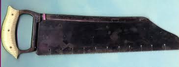
I'm sure you know how a saw sounds. Schrrck-schrrck-schrrck-schrrck. Usually we have one person sawing and the other four trying to hold the cadavre still. Sometimes we have everything dissected away and are right down on the bone, and sometimes we're just flat out sawing through the face or some such thing. That's always a little weird feeling.
A lot of other groups like to use chisels. I know this because you can hear a dozen chisels clinking away throughout the room. If you stop to listen, it sounds just like shop class. Chink, chink, schrrck-schrrck-schrrck, chink, clunk, clink, chink!
That said, what is it we're actually doing? As much as it might sound like it, we're not just tearing the face apart. The first sawing/chiseling we had to do was so we could get a good look at the jaw, and later we carefully took out a section of the muzzle, but the best was definitely when it was time to look at the brain. Despite having giant, unwieldy saws, we needed to plan and cut very carefully. You probably noticed that skulls are round, not square, and it's definitely not a good idea to start sawing into the brain. When we finally prised off the cranium, we learned that we had to then go hack off bone around the spinal cord so we could actually cut the brain stem and get the brain out. It was a long day.
At the end of the day, as normal and reasonable as it may have been at the time, it just does not do to try and tell your non-vet friends how you sawed a dog's face in half today.
Several times now, we've had to cut through bones in anatomy lab to get to what we want to look at. The first time was in the thorax, where we had to break off the ribs so we could look inside and see the heart and lungs and stuff. This involved using a pair of giant wire cutters ("bone cutters," sure) to snap the ribs at both ends with a loud crack. We left the muscles and everything in place, so that the thoracic wall makes a little flap that we can lift to peek into the thorax. How cute.
We only needed to go through bone one other time last semester, to see the spinal cord, and the rest of the bone sawing has been happening this semester. You see, this semester, we've spent seven weeks just looking at the head. There's a lot to see! Unfortunately a lot happens to be inside the head.
The thing that really made me want to tell a story about all this bone stuff is the noise. I already described the wire cutter bone cutters, but clearly that's not going to work very well for something like a skull. Our other options are saws and chisels.
My group likes to use the saw. It's completely barbaric feeling, but it gets the job done. The saws we have are these square things about as long as your forearm, that aren't too far off from this frightening picture I got from google:
I'm sure you know how a saw sounds. Schrrck-schrrck-schrrck-schrrck. Usually we have one person sawing and the other four trying to hold the cadavre still. Sometimes we have everything dissected away and are right down on the bone, and sometimes we're just flat out sawing through the face or some such thing. That's always a little weird feeling.
A lot of other groups like to use chisels. I know this because you can hear a dozen chisels clinking away throughout the room. If you stop to listen, it sounds just like shop class. Chink, chink, schrrck-schrrck-schrrck, chink, clunk, clink, chink!
That said, what is it we're actually doing? As much as it might sound like it, we're not just tearing the face apart. The first sawing/chiseling we had to do was so we could get a good look at the jaw, and later we carefully took out a section of the muzzle, but the best was definitely when it was time to look at the brain. Despite having giant, unwieldy saws, we needed to plan and cut very carefully. You probably noticed that skulls are round, not square, and it's definitely not a good idea to start sawing into the brain. When we finally prised off the cranium, we learned that we had to then go hack off bone around the spinal cord so we could actually cut the brain stem and get the brain out. It was a long day.
At the end of the day, as normal and reasonable as it may have been at the time, it just does not do to try and tell your non-vet friends how you sawed a dog's face in half today.
Thursday, 21 April 2011
Fuzzy Scalpels
I like scalpels, but they gave my anatomy group quite a bit of trouble when we were first starting out.
Sorry if this sounds creepy, but I love cutting with scalpels. It's so smooth! They're really sharp, so they make very nice, efficient cuts, and there's something satisfying about it, like a job well done.
So where does the trouble come in? Here is a picture that I got off google:

You have your metal handle, and the blade snaps on the top (the oblong thing you see in the middle of the blade is part of the handle). You can also see how where the bottom of the blade matches up with the handle, there's a diagonal line.
For starters, we keep putting the blades on backwards. Every time! The pointy part of the diagonal is always facing the wrong way. That's pretty embarrassing by itself, especially since it's been almost a year now. What makes that worse is that we had a very difficult time getting the blade back off at first.
You see, they had these little boxes that served as both a sharps container and an "easy" way to snap the blade off. You're supposed to stick the scalpel into a slot, twist it a bit, and the blade magically pops off. In theory. The first box we tried was so full, we couldn't even get the blade to go into the slot. The next two boxes we tried... well, the blade didn't magically pop off. Every single one of our lab group of five tried to jam it in and twist it around and finagle the blade off, but no one could get it to work. Eventually, we settled with just using forceps to pry the blade off, and hope it doesn't go flying or snap into pieces. I haven't seen the boxes around in a while, so I guess the faculty also decided they sucked and got rid of them.
Just as an ending note, why did I name the site Fuzzy Scalpel? Well the real reason is that I tried putting vet-related words together until I found two that I liked. But! (Warning, this may be a disturbing image for some) The image that came to mind was how when we cut through the skin of our dissection dogs, which are short haired dogs, the fur sticks all over the scalpel. This wouldn't happen in surgery because you shave and sterilise the operation site beforehand.
Saturday, 16 April 2011
Tail Wagging Dogs
To be sure that we took proper care of our dissection dogs, they started us off with a speech about showing respect. The anatomy professors pointed out how we should behave like professionals (for instance, not walk into the lab and go "Eeeeeeeeeeew smells disgusting!"). They explained how we only have one dog to last five people for the whole year, so if we didn't take care of it, we would be losing our learning material permanently. They described how much time and effort went into preparing the cadavres.
Because this is a pretty important point, it was reiterated often. Not too far into the semester, we were given a lecture about why these dogs deserved our respect. They were dogs that had to be euthanised due to society's failings. For example, a dog that had been bred for fighting and was rescued: it couldn't be re-homed because of the danger it might attack someone. The anatomy faculty stressed how they had personally been the ones to euthanise them, to see them as happy, tail wagging dogs, alive, and then how they had personally spent months of their time preserving them and making them suitable for our dissections (for example, filling the blood vessels with latex so we can identify arteries or veins).
This is a very touching concept and you can see why the dogs do deserve respect. Imagine having to put down a normal, cheerful dog, just because of a latent possibility of danger. Imagine pouring effort into preserving it, making its death worthwhile by advancing the learning of students. Then imagine some 19 year old waltzing into the lab, going up to your hard work, and complaining, "Eew it's disgusting!" Or imagine a group that didn't bother to store it properly, letting it to dry out or rot.
All that is very serious. However, the "happy, tail-wagging dog" spiel was recited to us over, and over, and over, and over. The first time, I thought it was pretty moving. Eventually it was like "Oh god, not again." Now, we often joke with each other, saying "You're being very unprofessional!" or "Show the dog some respect, it used to be a happy, tail-wagging dog!"
(This is of course in response to something said in jest, we do actually treat our dissection dog very well).
Because this is a pretty important point, it was reiterated often. Not too far into the semester, we were given a lecture about why these dogs deserved our respect. They were dogs that had to be euthanised due to society's failings. For example, a dog that had been bred for fighting and was rescued: it couldn't be re-homed because of the danger it might attack someone. The anatomy faculty stressed how they had personally been the ones to euthanise them, to see them as happy, tail wagging dogs, alive, and then how they had personally spent months of their time preserving them and making them suitable for our dissections (for example, filling the blood vessels with latex so we can identify arteries or veins).
This is a very touching concept and you can see why the dogs do deserve respect. Imagine having to put down a normal, cheerful dog, just because of a latent possibility of danger. Imagine pouring effort into preserving it, making its death worthwhile by advancing the learning of students. Then imagine some 19 year old waltzing into the lab, going up to your hard work, and complaining, "Eew it's disgusting!" Or imagine a group that didn't bother to store it properly, letting it to dry out or rot.
All that is very serious. However, the "happy, tail-wagging dog" spiel was recited to us over, and over, and over, and over. The first time, I thought it was pretty moving. Eventually it was like "Oh god, not again." Now, we often joke with each other, saying "You're being very unprofessional!" or "Show the dog some respect, it used to be a happy, tail-wagging dog!"
(This is of course in response to something said in jest, we do actually treat our dissection dog very well).
Thursday, 14 April 2011
Here is a dog. Have fun.
Our very first anatomy lab was not how I imagined it would be.
Beforehand, we had to pick our group of five that we would be stuck with for the entire year, but we didn't know each other very well so that developed very unfortunately for some people.
On the day of, we all showed up in our lab coats and clutching our lab manuals, hoping one person in our group had managed to get a dissection kit. Our anatomy lab is on the second floor, and there's two entrances as there are two stairways. At this point in time, I didn't know my way around the vet tower very well, and I went up the wrong stairway, so I was actually at the back entrance to the lab. I was getting worried, because out of a hundred people there was no one else waiting there, and only a few showed up behind me. I peeked into the room, but there definitely weren't students in there; I saw a big empty room and some serious-looking faculty talking to each other.
When it was finally time for lab, I crept in, and was relieved to see a massive flood of students coming from the other side of the room.
Now, you may not have thought about this, but to learn anatomy, vet students cut up dead dogs. Your doctors cut up dead people. This is a normal thing, the cadavres take months of preparation and hard work, and they are dogs that would have been euthanised one way or another. There's nothing inhumane about the dissection dogs. You just have to realise that the first thing you see on your first lab is a room of tables with dead dogs lying on their backs, their little paws sticking rigidly up in the air.
Apparently, a few years ago, someone went in after hours to do some review, and brought their non-vet friend with them. This person had never stopped to think about what would actually be in the anatomy lab, and freaked out when she saw the dead dogs. Her reaction? She went straight to the press. I think she thought that the school was covering up this big secret of slaughtering dogs and cutting them up, and they needed to be exposed. At any rate, nothing bad happened, because everything is regulated and has rules and things, but ever since then, no non-vet people are allowed in under any circumstances, and we can't take any pictures.
So here I am, faced with a sea of dead dogs, and we need to pick one that we'll be using for the next year. It's a big, wide room with drains along the floor, metal tables, stools, lots of sinks around the edges, and open windows covering three walls. It's very bright, there are TV monitors all over the room, and it smells like formaldehyde and disinfectant. I actually expected the smell to be much worse, but after enough labs it does start getting to you. Older students had told us to go for the Greyhounds because they have really nice muscles, but I wasn't fast enough to grab one. Since I came in through the back, there were a few skinny ones nearby, so I just picked one that looked sort of ok. I probably didn't make the best choice. For the record, having a fat dissection dog totally sucks. You have to get rid of the fat before you can see anything, and believe me the fat is everywhere.
The next step was to locate the rest of my lab group, who had been scratching their heads over some dogs on the other side of the room, get out the dissection kit, open the lab manuals, and figure out what to do.
This is where we ran into a problem. The faculty must not have been very organised because we didn't get much of a lecture on what to do or how to do it. You see, once we figured out how to get the blade on the scalpel, it was pretty much, "Go!" The lab manual was the only thing telling us what to do, but since we hadn't actually learned anatomy yet, the instructions were pretty much incomprehensible. Here are the first two sentences:
Looking back, the first few anatomy labs are pretty funny. Our labs now are very involved. This year we've done some insane stuff that is very memorable. Last year, we started out with very, very simple dissections, but at the time it was extremely important and complex to us. We would go really slowly and hesitantly, not sure whether we should cut something or not. The first labs compared to present labs are like cooking ramen noodles compared to Christmas dinner.
Beforehand, we had to pick our group of five that we would be stuck with for the entire year, but we didn't know each other very well so that developed very unfortunately for some people.
On the day of, we all showed up in our lab coats and clutching our lab manuals, hoping one person in our group had managed to get a dissection kit. Our anatomy lab is on the second floor, and there's two entrances as there are two stairways. At this point in time, I didn't know my way around the vet tower very well, and I went up the wrong stairway, so I was actually at the back entrance to the lab. I was getting worried, because out of a hundred people there was no one else waiting there, and only a few showed up behind me. I peeked into the room, but there definitely weren't students in there; I saw a big empty room and some serious-looking faculty talking to each other.
When it was finally time for lab, I crept in, and was relieved to see a massive flood of students coming from the other side of the room.
Now, you may not have thought about this, but to learn anatomy, vet students cut up dead dogs. Your doctors cut up dead people. This is a normal thing, the cadavres take months of preparation and hard work, and they are dogs that would have been euthanised one way or another. There's nothing inhumane about the dissection dogs. You just have to realise that the first thing you see on your first lab is a room of tables with dead dogs lying on their backs, their little paws sticking rigidly up in the air.
Apparently, a few years ago, someone went in after hours to do some review, and brought their non-vet friend with them. This person had never stopped to think about what would actually be in the anatomy lab, and freaked out when she saw the dead dogs. Her reaction? She went straight to the press. I think she thought that the school was covering up this big secret of slaughtering dogs and cutting them up, and they needed to be exposed. At any rate, nothing bad happened, because everything is regulated and has rules and things, but ever since then, no non-vet people are allowed in under any circumstances, and we can't take any pictures.
So here I am, faced with a sea of dead dogs, and we need to pick one that we'll be using for the next year. It's a big, wide room with drains along the floor, metal tables, stools, lots of sinks around the edges, and open windows covering three walls. It's very bright, there are TV monitors all over the room, and it smells like formaldehyde and disinfectant. I actually expected the smell to be much worse, but after enough labs it does start getting to you. Older students had told us to go for the Greyhounds because they have really nice muscles, but I wasn't fast enough to grab one. Since I came in through the back, there were a few skinny ones nearby, so I just picked one that looked sort of ok. I probably didn't make the best choice. For the record, having a fat dissection dog totally sucks. You have to get rid of the fat before you can see anything, and believe me the fat is everywhere.
The next step was to locate the rest of my lab group, who had been scratching their heads over some dogs on the other side of the room, get out the dissection kit, open the lab manuals, and figure out what to do.
This is where we ran into a problem. The faculty must not have been very organised because we didn't get much of a lecture on what to do or how to do it. You see, once we figured out how to get the blade on the scalpel, it was pretty much, "Go!" The lab manual was the only thing telling us what to do, but since we hadn't actually learned anatomy yet, the instructions were pretty much incomprehensible. Here are the first two sentences:
"Make a median ventral incision from the middle of the neck to the xiphoid cartilage. From a point of this incision opposite the brachium, extend a transverse incision to the elbow on one side."Good luck.
Looking back, the first few anatomy labs are pretty funny. Our labs now are very involved. This year we've done some insane stuff that is very memorable. Last year, we started out with very, very simple dissections, but at the time it was extremely important and complex to us. We would go really slowly and hesitantly, not sure whether we should cut something or not. The first labs compared to present labs are like cooking ramen noodles compared to Christmas dinner.
Subscribe to:
Posts (Atom)