You might be wondering how surgeons get inside the turtle. It turns out that one of the methods is to make an incision in the soft area in front of their back legs (or front, depending on what you want to do). Then they can stick an endoscope in, with a light source and a camera on it, and use tiny instruments with long handles. The best part is that the light makes the internal cavity of the turtle glow, and you can see it through the shell on their belly!
I can't find any good photos on google, so I'm going to show you the ones I took. I'm not sure if we're allowed to do that, so uh, don't tell the school on me please.

Intubated and with an IV catheter in her jugular.

The reason the turtle looks so yellow is because there's a sticky sterile covering with iodine in it, called Ioban.

If you look carefully, you can see how bright the pink inside of the turtle is because of the light from the endoscope.
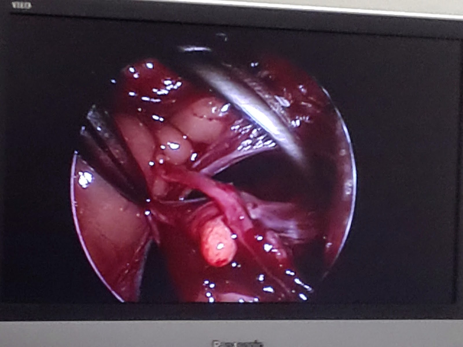
The screen showing the instruments and tissues on the endoscope.
Why was this turtle having surgery? The short and simple version is that it had egg impaction. And how did they definitively diagnose that? A CT scan. In case you were wondering, here's a picture from google of a turtle CT scan:

No comments:
Post a Comment